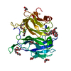
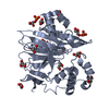
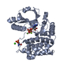
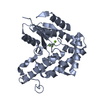
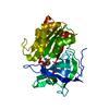
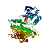
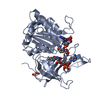
Entry Database : PDB / ID : 5jebTitle Crystal structure of EGFR tyrosine kinase domain with novel inhibitor of active state of HER2 Epidermal growth factor receptor Keywords / / / Function / homology Function Domain/homology Component
/ / / / / / / / / / / / / / / / / / / / / / / / / / / / / / / / / / / / / / / / / / / / / / / / / / / / / / / / / / / / / / / / / / / / / / / / / / / / / / / / / / / / / / / / / / / / / / / / / / / / / / / / / / / / / / / / / / / / / / / / / / / / / / / / / / / / / / / / / / / / / / / / / / / / / Biological species Homo sapiens (human)Method / / / Resolution : 3.298 Å Authors Park, J.H. / Lemmon, M.A. Funding support Organization Grant number Country National Institutes of Health/National Institute of General Medical Sciences (NIH/NIGMS) R01-GM099891
Journal : Nat.Chem.Biol. / Year : 2016Title : Overcoming resistance to HER2 inhibitors through state-specific kinase binding.Authors : Novotny, C.J. / Pollari, S. / Park, J.H. / Lemmon, M.A. / Shen, W. / Shokat, K.M. History Deposition Apr 18, 2016 Deposition site / Processing site Revision 1.0 Sep 7, 2016 Provider / Type Revision 1.1 Sep 21, 2016 Group Revision 1.2 Nov 2, 2016 Group Revision 1.3 Sep 27, 2017 Group / Author supporting evidence / Derived calculationsCategory / pdbx_struct_oper_list / pdbx_unobs_or_zero_occ_atomsItem / _pdbx_struct_oper_list.symmetry_operationRevision 1.4 Dec 25, 2019 Group / Category / Item Revision 1.5 Sep 27, 2023 Group Advisory / Data collection ... Advisory / Data collection / Database references / Refinement description Category chem_comp_atom / chem_comp_bond ... chem_comp_atom / chem_comp_bond / database_2 / pdbx_initial_refinement_model / pdbx_unobs_or_zero_occ_atoms Item / _database_2.pdbx_database_accession
Show all Show less
 Movie
Movie Controller

 Yorodumi
 Open data
 Basic information
 Components
 Keywords
 Function and homology information
 Homo sapiens (human)
 X-RAY DIFFRACTION /
  Authors
 United States, 1items
  Citation
 Structure visualization
 Downloads & links
 Other downloads
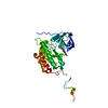


 PDBj
PDBj


 Assembly
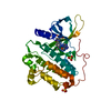

 Spodoptera frugiperda (fall armyworm)
 Mass: 96.063 Da / Num. of mol.: 2 / Source method: obtained synthetically / Formula: SO4
 Mass: 373.365 Da / Num. of mol.: 1 / Source method: obtained synthetically / Formula: C20H15N5O3
 Sample preparation
 Processing