Protocol: SINGLE WAVELENGTH / Monochromatic (M) / Laue (L): M / Scattering type: x-ray
Radiation wavelength
Wavelength: 1 Å / Relative weight: 1
Reflection
Resolution: 1.14→42.86 Å / Num. obs: 44583 / % possible obs: 97.9 % / Redundancy: 6.16 % / Biso Wilson estimate: 16.393 Å2 / CC1/2: 0.999 / Rmerge(I) obs: 0.0658 / Net I/σ(I): 10.27
Reflection shell
Resolution: 1.14→1.24 Å / Redundancy: 5.71 % / Rmerge(I) obs: 0.6068 / Mean I/σ(I) obs: 2.04 / Rsym value: 0.6068 / % possible all: 94.3
-
Processing
Software
Name
Version
Classification
XSCALE
datascaling
PDB_EXTRACT
3.2
dataextraction
REFMAC
5.6.0112
refinement
XDS
datareduction
REFMAC
phasing
Refinement
Method to determine structure: MOLECULAR REPLACEMENT Starting model: IN-HOUSE STRUCTURE Resolution: 1.14→42.86 Å / Cor.coef. Fo:Fc: 0.981 / Cor.coef. Fo:Fc free: 0.973 / SU B: 1.261 / SU ML: 0.026 / Cross valid method: FREE R-VALUE / ESU R: 0.034 / ESU R Free: 0.037 / Stereochemistry target values: MAXIMUM LIKELIHOOD Details: HYDROGENS HAVE BEEN USED IF PRESENT IN THE INPUT. U VALUES REFINED INDIVIDUALLY
Rfactor
Num. reflection
% reflection
Selection details
Rfree
0.17342
2183
5.1 %
RANDOM
Rwork
0.1358
-
-
-
obs
0.13772
41025
95 %
-
Solvent computation
Ion probe radii: 0.8 Å / Shrinkage radii: 0.8 Å / VDW probe radii: 1.2 Å / Solvent model: BABINET MODEL WITH MASK
 Movie
Movie Controller

 Yorodumi
 Open data
 Basic information
 Components
 Keywords
 Function and homology information
 Homo sapiens (human)
 X-RAY DIFFRACTION /
  Authors
 Citation
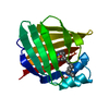
 Structure visualization
 Downloads & links
 Other downloads


 PDBj
PDBj

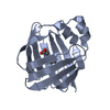
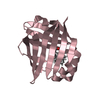
 Assembly
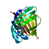

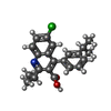
 Mass: 367.869 Da / Num. of mol.: 1 / Source method: obtained synthetically / Formula: C22H22ClNO2
 Mass: 18.015 Da / Num. of mol.: 226 / Source method: isolated from a natural source / Formula: H2O
 Sample preparation
 / Beamline: X10SA / Wavelength: 1 Å
 Processing