 Movie
Movie Controller

[English] 日本語
 Yorodumi
- PDB-5ezh: Structure of Transcriptional Regulatory Repressor Protein - EthR ... -
+ Open data

- Basic information
| Entry | Database: PDB / ID: 5ezh | ||||||||||||
|---|---|---|---|---|---|---|---|---|---|---|---|---|---|
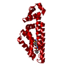
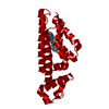
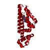
| Title | Structure of Transcriptional Regulatory Repressor Protein - EthR from Mycobacterium Tuberculosis in complex with compound 21 at 1.7A resolution | ||||||||||||
 Components Components | HTH-type transcriptional regulator EthR | ||||||||||||
 Keywords Keywords | TRANSCRIPTION / EthR / repressor / Mycobacterium tuberculosis | ||||||||||||
| Function / homology |  Function and homology information Function and homology informationtranscription cis-regulatory region binding / DNA-binding transcription factor activity / response to antibiotic / negative regulation of DNA-templated transcription / regulation of DNA-templated transcription / cytosol Similarity search - Function | ||||||||||||
| Biological species |   Mycobacterium tuberculosis (bacteria) Mycobacterium tuberculosis (bacteria) | ||||||||||||
| Method |  X-RAY DIFFRACTION / SYNCHROTRON / MOLECULAR REPLACEMENT / molecular replacement / Resolution: 1.7 Å X-RAY DIFFRACTION / SYNCHROTRON / MOLECULAR REPLACEMENT / molecular replacement / Resolution: 1.7 Å | ||||||||||||
 Authors Authors | Surade, S. / Blaszczyk, M. / Nikiforov, P.O. / Abell, C. / Blundell, T.L. | ||||||||||||
| Funding support |  United Kingdom, United Kingdom,  United States, 3items United States, 3items
| ||||||||||||
 Citation Citation | Journal: Org.Biomol.Chem. / Year: 2016 Title: A fragment merging approach towards the development of small molecule inhibitors of Mycobacterium tuberculosis EthR for use as ethionamide boosters. Authors: Nikiforov, P.O. / Surade, S. / Blaszczyk, M. / Delorme, V. / Brodin, P. / Baulard, A.R. / Blundell, T.L. / Abell, C. | ||||||||||||
| History |
|
- Structure visualization
| Structure viewer | Molecule: MolmilJmol/JSmol |
|---|
- Downloads & links
-Download
| PDBx/mmCIF format | 5ezh.cif.gz | 55.1 KB | Display | PDBx/mmCIF format |
|---|---|---|---|---|
| PDB format | pdb5ezh.ent.gz | 38.3 KB | Display | PDB format |
| PDBx/mmJSON format | 5ezh.json.gz | Tree view | PDBx/mmJSON format | |
| Others |  Other downloads Other downloads |
-Validation report
| Summary document | 5ezh_validation.pdf.gz | 956.5 KB | Display | wwPDB validaton report |
|---|---|---|---|---|
| Full document | 5ezh_full_validation.pdf.gz | 959 KB | Display | |
| Data in XML | 5ezh_validation.xml.gz | 10.4 KB | Display | |
| Data in CIF | 5ezh_validation.cif.gz | 13.7 KB | Display | |
| Arichive directory | https://data.pdbj.org/pub/pdb/validation_reports/ez/5ezhftp://data.pdbj.org/pub/pdb/validation_reports/ez/5ezh | HTTPS FTP |
-Related structure data
| Related structure data | 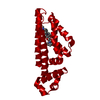 5eyrC 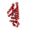 5ezgC 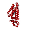 5f04C 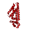 5f08C  5f0cC  5f0fC  5f0hC  5f1jC 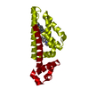 5f27C 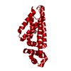 1t56S S: Starting model for refinement C: citing same article ( |
|---|---|
| Similar structure data |


-Links
 PDBj
PDBj- Assembly
| Deposited unit | 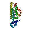
| |||||||||
|---|---|---|---|---|---|---|---|---|---|---|
| 1 | 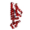
| |||||||||
| Unit cell |
| |||||||||
| Components on special symmetry positions |
|
-Components
| #1: Protein | Mass: 25259.254 Da / Num. of mol.: 1 Source method: isolated from a genetically manipulated source Source: (gene. exp.) Mycobacterium tuberculosis (bacteria) / Gene: ethR, etaR, MT3970 / Production host: | ||||
|---|---|---|---|---|---|
| #2: Chemical |   Mass: 288.388 Da / Num. of mol.: 2 / Source method: obtained synthetically / Formula: C16H24N4O Mass: 288.388 Da / Num. of mol.: 2 / Source method: obtained synthetically / Formula: C16H24N4O#3: Chemical | ChemComp-SO4 / |   Mass: 96.063 Da / Num. of mol.: 1 / Source method: obtained synthetically / Formula: SO4 Mass: 96.063 Da / Num. of mol.: 1 / Source method: obtained synthetically / Formula: SO4#4: Water | ChemComp-HOH / |  Mass: 18.015 Da / Num. of mol.: 49 / Source method: isolated from a natural source / Formula: H2O Mass: 18.015 Da / Num. of mol.: 49 / Source method: isolated from a natural source / Formula: H2O |
-Experimental details
-Experiment
| Experiment | Method: X-RAY DIFFRACTION / Number of used crystals: 1 |
|---|
- Sample preparation
| Crystal | Density Matthews: 2.47 Å3/Da / Density % sol: 50.25 % |
|---|---|
| Crystal grow | Temperature: 298 K / Method: vapor diffusion, sitting drop / Details: Ammonium sulphate, Glycerol, MES / PH range: 6.3 - 6.5 |
-Data collection
| Diffraction | Mean temperature: 100 K | |||||||||||||||||||||||||||
|---|---|---|---|---|---|---|---|---|---|---|---|---|---|---|---|---|---|---|---|---|---|---|---|---|---|---|---|---|
| Diffraction source | Source: SYNCHROTRON / Site: Diamond / Beamline: I04 / Wavelength: 0.97943 Å | |||||||||||||||||||||||||||
| Detector | Type: DECTRIS PILATUS 6M / Detector: PIXEL / Date: Dec 11, 2013 | |||||||||||||||||||||||||||
| Radiation | Protocol: SINGLE WAVELENGTH / Monochromatic (M) / Laue (L): M / Scattering type: x-ray | |||||||||||||||||||||||||||
| Radiation wavelength | Wavelength: 0.97943 Å / Relative weight: 1 | |||||||||||||||||||||||||||
| Reflection | Resolution: 1.7→85.85 Å / Num. obs: 28584 / % possible obs: 100 % / Redundancy: 12.6 % / CC1/2: 1 / Rmerge(I) obs: 0.051 / Rpim(I) all: 0.015 / Net I/σ(I): 28.4 / Num. measured all: 359919 | |||||||||||||||||||||||||||
| Reflection shell | Diffraction-ID: 1 / Rejects: _
|
-Phasing
| Phasing | Method: molecular replacement | |||||||||
|---|---|---|---|---|---|---|---|---|---|---|
| Phasing MR | Model details: Phaser MODE: MR_AUTO
|
- Processing
| Software |
| |||||||||||||||||||||||||||||||||||||||||||||
|---|---|---|---|---|---|---|---|---|---|---|---|---|---|---|---|---|---|---|---|---|---|---|---|---|---|---|---|---|---|---|---|---|---|---|---|---|---|---|---|---|---|---|---|---|---|---|
| Refinement | Method to determine structure: MOLECULAR REPLACEMENT Starting model: 1T56 Resolution: 1.7→60.71 Å / Cor.coef. Fo:Fc: 0.959 / Cor.coef. Fo:Fc free: 0.945 / WRfactor Rfree: 0.2302 / WRfactor Rwork: 0.1884 / FOM work R set: 0.8655 / SU B: 1.671 / SU ML: 0.057 / SU R Cruickshank DPI: 0.093 / SU Rfree: 0.0974 / Cross valid method: THROUGHOUT / σ(F): 0 / ESU R: 0.093 / ESU R Free: 0.097 / Stereochemistry target values: MAXIMUM LIKELIHOOD Details: HYDROGENS HAVE BEEN USED IF PRESENT IN THE INPUT U VALUES : REFINED INDIVIDUALLY
| |||||||||||||||||||||||||||||||||||||||||||||
| Solvent computation | Ion probe radii: 0.8 Å / Shrinkage radii: 0.8 Å / VDW probe radii: 1.2 Å / Solvent model: MASK | |||||||||||||||||||||||||||||||||||||||||||||
| Displacement parameters | Biso max: 78.27 Å2 / Biso mean: 28.56 Å2 / Biso min: 12.19 Å2
| |||||||||||||||||||||||||||||||||||||||||||||
| Refinement step | Cycle: final / Resolution: 1.7→60.71 Å
| |||||||||||||||||||||||||||||||||||||||||||||
| Refine LS restraints |
| |||||||||||||||||||||||||||||||||||||||||||||
| LS refinement shell | Resolution: 1.7→1.744 Å / Total num. of bins used: 20
|
