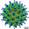
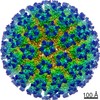
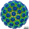
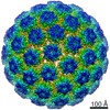
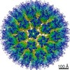
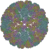
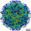
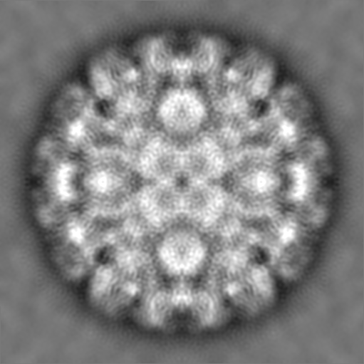
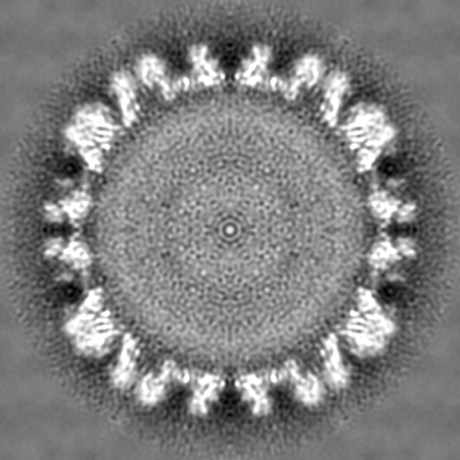
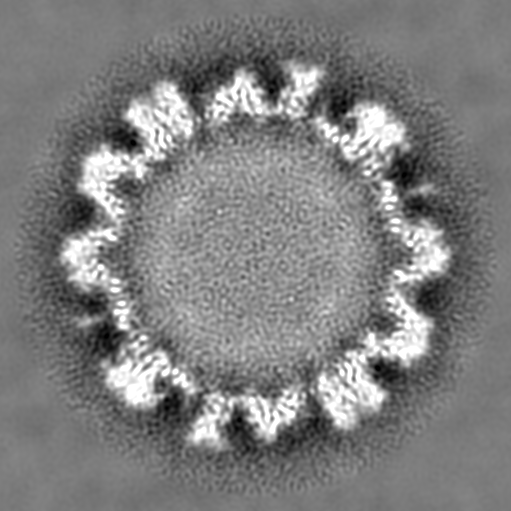
- EMDB-5932: Electron cryo-microscopy of Human Papillomavirus Type 16 Capsid -
+
データを開く
IDまたはキーワード:
読み込み中...
-
基本情報
登録情報
データベース: EMDB / ID: EMD-5932
タイトル
Electron cryo-microscopy of Human Papillomavirus Type 16 Capsid
マップデータ
Reconstruction of fully mature HPV16 L1/L2 capsid
試料
試料: Human Papilloma Virus type 16 capsid
ウイルス: Human papillomavirus type 16 (パピローマウイルス)
機能・相同性
機能・相同性情報
T=7 icosahedral viral capsid / endocytosis involved in viral entry into host cell / host cell nucleus / virion attachment to host cell / structural molecule activity 類似検索 - 分子機能
Major capsid L1 (late) protein, Papillomavirus / Major capsid L1 (late) superfamily, Papillomavirus / L1 (late) protein / Double-stranded DNA virus, group I, capsid 類似検索 - ドメイン・相同性
Major capsid protein L1 / Major capsid protein L1 類似検索 - 構成要素
ジャーナル: mBio / 年: 2014 タイトル: Maturation of the human papillomavirus 16 capsid. 著者: Giovanni Cardone / Adam L Moyer / Naiqian Cheng / Cynthia D Thompson / Israel Dvoretzky / Douglas R Lowy / John T Schiller / Alasdair C Steven / Christopher B Buck / Benes L Trus / 要旨: Papillomaviruses are a family of nonenveloped DNA viruses that infect the skin or mucosa of their vertebrate hosts. The viral life cycle is closely tied to the differentiation of infected ...Papillomaviruses are a family of nonenveloped DNA viruses that infect the skin or mucosa of their vertebrate hosts. The viral life cycle is closely tied to the differentiation of infected keratinocytes. Papillomavirus virions are released into the environment through a process known as desquamation, in which keratinocytes lose structural integrity prior to being shed from the surface of the skin. During this process, virions are exposed to an increasingly oxidative environment, leading to their stabilization through the formation of disulfide cross-links between neighboring molecules of the major capsid protein, L1. We used time-lapse cryo-electron microscopy and image analysis to study the maturation of HPV16 capsids assembled in mammalian cells and exposed to an oxidizing environment after cell lysis. Initially, the virion is a loosely connected procapsid that, under in vitro conditions, condenses over several hours into the more familiar 60-nm-diameter papillomavirus capsid. In this process, the procapsid shrinks by ~5% in diameter, its pentameric capsomers change in structure (most markedly in the axial region), and the interaction surfaces between adjacent capsomers are consolidated. A C175S mutant that cannot achieve normal inter-L1 disulfide cross-links shows maturation-related shrinkage but does not achieve the fully condensed 60-nm form. Pseudoatomic modeling based on a 9-Å resolution reconstruction of fully mature capsids revealed C-terminal disulfide-stabilized "suspended bridges" that form intercapsomeric cross-links. The data suggest a model in which procapsids exist in a range of dynamic intermediates that can be locked into increasingly mature configurations by disulfide cross-linking, possibly through a Brownian ratchet mechanism. Importance: Human papillomaviruses (HPVs) cause nearly all cases of cervical cancer, a major fraction of cancers of the penis, vagina/vulva, anus, and tonsils, and genital and nongenital warts. HPV types associated with a high risk of cancer, such as HPV16, are generally transmitted via sexual contact. The nonenveloped virion of HPVs shows a high degree of stability, allowing the virus to persist in an infectious form in environmental fomites. In this study, we used cryo-electron microscopy to elucidate the structure of the HPV16 capsid at different stages of maturation. The fully mature capsid adopts a rigid, highly regular structure stabilized by intermolecular disulfide bonds. The availability of a pseudoatomic model of the fully mature HPV16 virion should help guide understanding of antibody responses elicited by HPV capsid-based vaccines.
 ムービー
ムービー コントローラー


 データを開く
 基本情報
基本情報 マップデータ
 試料
試料 機能・相同性情報
 Human papillomavirus type 16 (パピローマウイルス)
 データ登録者
 引用
引用
 構造の表示
構造の表示
 ダウンロードとリンク
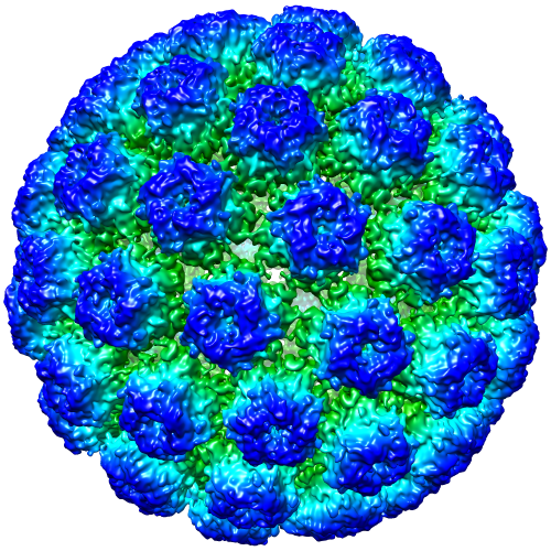 emd_5932.png
 http://ftp.pdbj.org/pub/emdb/structures/EMD-5932
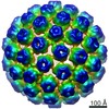


 Z (Sec.)
 Y (Row.)
 X (Col.)


 試料の構成要素
 Homo sapiens (ヒト) / 別称: VERTEBRATES
 解析
解析 電子顕微鏡法
 FIELD EMISSION GUN

