+ データを開く
- 基本情報
基本情報
| 登録情報 | データベース: EMDB / ID: EMD-5527 | |||||||||
|---|---|---|---|---|---|---|---|---|---|---|
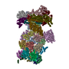
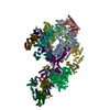
| タイトル | Reconstruction of the deltaGCC mutant HCV IRES bound to rabbit 40S ribosomal subunit | |||||||||
 マップデータ マップデータ | Reconstruction of mutant HCV IRES bound to 40S | |||||||||
 試料 試料 |
| |||||||||
 キーワード キーワード | HCV IRES / 40S | |||||||||
| 生物種 |   Hepatitis C virus (ウイルス) Hepatitis C virus (ウイルス) | |||||||||
| 手法 | 単粒子再構成法 / クライオ電子顕微鏡法 / 解像度: 17.5 Å | |||||||||
 データ登録者 データ登録者 | Vollmar BS / Shi D / Filbin ME / Kieft JS / Gonen T | |||||||||
 引用 引用 |  ジャーナル: Nat Struct Mol Biol / 年: 2013 ジャーナル: Nat Struct Mol Biol / 年: 2013タイトル: HCV IRES manipulates the ribosome to promote the switch from translation initiation to elongation. 著者: Megan E Filbin / Breanna S Vollmar / Dan Shi / Tamir Gonen / Jeffrey S Kieft /  要旨: The internal ribosome entry site (IRES) of the hepatitis C virus (HCV) drives noncanonical initiation of protein synthesis necessary for viral replication. Functional studies of the HCV IRES have ...The internal ribosome entry site (IRES) of the hepatitis C virus (HCV) drives noncanonical initiation of protein synthesis necessary for viral replication. Functional studies of the HCV IRES have focused on 80S ribosome formation but have not explored its role after the 80S ribosome is poised at the start codon. Here, we report that mutations of an IRES domain that docks in the 40S subunit's decoding groove cause only a local perturbation in IRES structure and result in conformational changes in the IRES-rabbit 40S subunit complex. Functionally, the mutations decrease IRES activity by inhibiting the first ribosomal translocation event, and modeling results suggest that this effect occurs through an interaction with a single ribosomal protein. The ability of the HCV IRES to manipulate the ribosome provides insight into how the ribosome's structure and function can be altered by bound RNAs, including those derived from cellular invaders. | |||||||||
| 履歴 |
|
- 構造の表示
構造の表示
| ムービー |
 ムービービューア ムービービューア |
|---|---|
| 構造ビューア | EMマップ:  SurfView SurfView Molmil Molmil Jmol/JSmol Jmol/JSmol |
| 添付画像 |
- ダウンロードとリンク
-EMDBアーカイブ
| マップデータ |  emd_5527.map.gz emd_5527.map.gz | 11.1 MB |  EMDBマップデータ形式 EMDBマップデータ形式 | |
|---|---|---|---|---|
| ヘッダ (付随情報) |  emd-5527-v30.xml emd-5527-v30.xml emd-5527.xml emd-5527.xml | 9.2 KB 9.2 KB | 表示 表示 |  EMDBヘッダ EMDBヘッダ |
| 画像 |  emd_5527_1.tif emd_5527_1.tif | 164.9 KB | ||
| アーカイブディレクトリ |  http://ftp.pdbj.org/pub/emdb/structures/EMD-5527 http://ftp.pdbj.org/pub/emdb/structures/EMD-5527 ftp://ftp.pdbj.org/pub/emdb/structures/EMD-5527 ftp://ftp.pdbj.org/pub/emdb/structures/EMD-5527 | HTTPS FTP |
-検証レポート
| 文書・要旨 |  emd_5527_validation.pdf.gz emd_5527_validation.pdf.gz | 77.9 KB | 表示 |  EMDB検証レポート EMDB検証レポート |
|---|---|---|---|---|
| 文書・詳細版 |  emd_5527_full_validation.pdf.gz emd_5527_full_validation.pdf.gz | 77 KB | 表示 | |
| XML形式データ |  emd_5527_validation.xml.gz emd_5527_validation.xml.gz | 493 B | 表示 | |
| アーカイブディレクトリ |  https://ftp.pdbj.org/pub/emdb/validation_reports/EMD-5527 https://ftp.pdbj.org/pub/emdb/validation_reports/EMD-5527 ftp://ftp.pdbj.org/pub/emdb/validation_reports/EMD-5527 ftp://ftp.pdbj.org/pub/emdb/validation_reports/EMD-5527 | HTTPS FTP |
-関連構造データ
- リンク
リンク
| EMDBのページ |  EMDB (EBI/PDBe) / EMDB (EBI/PDBe) /  EMDataResource EMDataResource |
|---|---|
| 「今月の分子」の関連する項目 |
- マップ
マップ
| ファイル |  ダウンロード / ファイル: emd_5527.map.gz / 形式: CCP4 / 大きさ: 12.6 MB / タイプ: IMAGE STORED AS FLOATING POINT NUMBER (4 BYTES) ダウンロード / ファイル: emd_5527.map.gz / 形式: CCP4 / 大きさ: 12.6 MB / タイプ: IMAGE STORED AS FLOATING POINT NUMBER (4 BYTES) | ||||||||||||||||||||||||||||||||||||||||||||||||||||||||||||||||||||
|---|---|---|---|---|---|---|---|---|---|---|---|---|---|---|---|---|---|---|---|---|---|---|---|---|---|---|---|---|---|---|---|---|---|---|---|---|---|---|---|---|---|---|---|---|---|---|---|---|---|---|---|---|---|---|---|---|---|---|---|---|---|---|---|---|---|---|---|---|---|
| 注釈 | Reconstruction of mutant HCV IRES bound to 40S | ||||||||||||||||||||||||||||||||||||||||||||||||||||||||||||||||||||
| 投影像・断面図 | 画像のコントロール
画像は Spider により作成 | ||||||||||||||||||||||||||||||||||||||||||||||||||||||||||||||||||||
| ボクセルのサイズ | X=Y=Z: 2.66 Å | ||||||||||||||||||||||||||||||||||||||||||||||||||||||||||||||||||||
| 密度 |
| ||||||||||||||||||||||||||||||||||||||||||||||||||||||||||||||||||||
| 対称性 | 空間群: 1 | ||||||||||||||||||||||||||||||||||||||||||||||||||||||||||||||||||||
| 詳細 | EMDB XML:
CCP4マップ ヘッダ情報:
| ||||||||||||||||||||||||||||||||||||||||||||||||||||||||||||||||||||
-添付データ
- 試料の構成要素
-全体 : mutant HCV IRES bound to 40S
| 全体 | 名称: mutant HCV IRES bound to 40S |
|---|---|
| 要素 |
|
-超分子 #1000: mutant HCV IRES bound to 40S
| 超分子 | 名称: mutant HCV IRES bound to 40S / タイプ: sample / ID: 1000 / Number unique components: 2 |
|---|
-超分子 #1: Rabbit 40S ribosomal subunit
| 超分子 | 名称: Rabbit 40S ribosomal subunit / タイプ: complex / ID: 1 / Name.synonym: 40S ribosome / 詳細: Mutant HCV IRES bound to 40S / 組換発現: No / データベース: NCBI / Ribosome-details: ribosome-eukaryote: SSU 40S |
|---|---|
| 由来(天然) | 生物種:  |
-分子 #1: hepatitis C virus internal ribosome entry site
| 分子 | 名称: hepatitis C virus internal ribosome entry site / タイプ: rna / ID: 1 / Name.synonym: HCV IRES 詳細: Deletion of GCC (82-84) of HCV IRES (nucleotides 40-372) 分類: OTHER / Structure: DOUBLE HELIX / Synthetic?: Yes |
|---|---|
| 由来(天然) | 生物種:  Hepatitis C virus (ウイルス) / 別称: HCV Hepatitis C virus (ウイルス) / 別称: HCV |
-実験情報
-構造解析
| 手法 | クライオ電子顕微鏡法 |
|---|---|
 解析 解析 | 単粒子再構成法 |
| 試料の集合状態 | particle |
- 試料調製
試料調製
| 緩衝液 | pH: 7.4 詳細: 20 mM Tris-HCl, pH 7.4, 200 mM KOAc, 2.5 mM MgCl2, 2 mM DTT, 40 mM KCl |
|---|---|
| グリッド | 詳細: Quantifoil R1.2/1.3 400 mesh copper grids with holey carbon |
| 凍結 | 凍結剤: ETHANE / チャンバー内湿度: 100 % / 装置: FEI VITROBOT MARK IV / 手法: Blot and plunge into liquid ethane |
- 電子顕微鏡法
| 顕微鏡 | FEI TECNAI F20 |
|---|---|
| 日付 | 2012年3月19日 |
| 撮影 | カテゴリ: CCD フィルム・検出器のモデル: GENERIC TVIPS (4k x 4k) 実像数: 1790 / 平均電子線量: 15 e/Å2 詳細: Used 2x binned images yielding a pixel size of 2.66 A/pixel. |
| Tilt angle min | 0 |
| Tilt angle max | 0 |
| 電子線 | 加速電圧: 200 kV / 電子線源:  FIELD EMISSION GUN FIELD EMISSION GUN |
| 電子光学系 | 倍率(補正後): 117293 / 照射モード: SPOT SCAN / 撮影モード: BRIGHT FIELD / Cs: 2.0 mm / 最大 デフォーカス(公称値): 5.0 µm / 最小 デフォーカス(公称値): 1.25 µm / 倍率(公称値): 62000 |
| 試料ステージ | 試料ホルダーモデル: GATAN LIQUID NITROGEN |
| 実験機器 |  モデル: Tecnai F20 / 画像提供: FEI Company |
- 画像解析
画像解析
| 詳細 | Particles selected manually using Electron Micrographe Utility (cryoem.ucsf.edu) |
|---|---|
| CTF補正 | 詳細: CTFFIND3 |
| 最終 再構成 | 解像度のタイプ: BY AUTHOR / 解像度: 17.5 Å / 解像度の算出法: OTHER / ソフトウェア - 名称: FREALIGN / 使用した粒子像数: 29920 |
 ムービー
ムービー コントローラー


 Z (Sec.)
 Y (Row.)
 X (Col.)


