+ データを開く
- 基本情報
基本情報
| 登録情報 | データベース: EMDB / ID: EMD-5116 | |||||||||
|---|---|---|---|---|---|---|---|---|---|---|
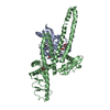
| タイトル | 30S subunit of ribosomal protein S1 | |||||||||
 マップデータ マップデータ | surface view of S1 protein | |||||||||
 試料 試料 |
| |||||||||
 キーワード キーワード | protein S1 / EM density / spider / ribosome / single particle reconstruction | |||||||||
| 生物種 | unidentified (未定義) | |||||||||
| 手法 | 単粒子再構成法 / クライオ電子顕微鏡法 / 解像度: 11.5 Å | |||||||||
 データ登録者 データ登録者 | Sengupta J / Agrawal RK / Frank J | |||||||||
 引用 引用 |  ジャーナル: Proc Natl Acad Sci U S A / 年: 2001 ジャーナル: Proc Natl Acad Sci U S A / 年: 2001タイトル: Visualization of protein S1 within the 30S ribosomal subunit and its interaction with messenger RNA. 著者: J Sengupta / R K Agrawal / J Frank /  要旨: S1 is the largest ribosomal protein, present in the small subunit of the bacterial ribosome. It has a pivotal role in stabilizing the mRNA on the ribosome. Thus far, S1 has eluded structural ...S1 is the largest ribosomal protein, present in the small subunit of the bacterial ribosome. It has a pivotal role in stabilizing the mRNA on the ribosome. Thus far, S1 has eluded structural determination. We have identified the S1 protein mass in the cryo-electron microscopic map of the Escherichia coli ribosome by comparing the map with a recent x-ray crystallographic structure of the 30S subunit, which lacks S1. According to our finding, S1 is located at the junction of head, platform, and main body of the 30S subunit, thus explaining all existing biochemical and crosslinking data. Protein S1 as identified in our map has a complex, elongated shape with two holes in its central portion. The N-terminal domain, forming one of the extensions, penetrates into the head of the 30S subunit. Evidence for direct interaction of S1 with 11 nucleotides of the mRNA, immediately upstream of the Shine-Dalgarno sequence, explains the protein's role in the recognition of the 5' region of mRNA. #1:  ジャーナル: CELL (CAMBRIDGE,MASS.) / 年: 2000 ジャーナル: CELL (CAMBRIDGE,MASS.) / 年: 2000タイトル: Solution structure of the E. coli 70S ribosome at 11.5 angstrom resolution 著者: Gabashvili IS / Agrawal RK / Spahn CMT / Grassucci RA / Svergun DI / Frank J / Penczek P | |||||||||
| 履歴 |
|
- 構造の表示
構造の表示
| ムービー |
 ムービービューア ムービービューア |
|---|---|
| 構造ビューア | EMマップ:  SurfView SurfView Molmil Molmil Jmol/JSmol Jmol/JSmol |
| 添付画像 |
- ダウンロードとリンク
-EMDBアーカイブ
| マップデータ |  emd_5116.map.gz emd_5116.map.gz | 7 MB |  EMDBマップデータ形式 EMDBマップデータ形式 | |
|---|---|---|---|---|
| ヘッダ (付随情報) |  emd-5116-v30.xml emd-5116-v30.xml emd-5116.xml emd-5116.xml | 7.7 KB 7.7 KB | 表示 表示 |  EMDBヘッダ EMDBヘッダ |
| 画像 |  emd_5116_1.png emd_5116_1.png | 101.4 KB | ||
| アーカイブディレクトリ |  http://ftp.pdbj.org/pub/emdb/structures/EMD-5116 http://ftp.pdbj.org/pub/emdb/structures/EMD-5116 ftp://ftp.pdbj.org/pub/emdb/structures/EMD-5116 ftp://ftp.pdbj.org/pub/emdb/structures/EMD-5116 | HTTPS FTP |
-検証レポート
| 文書・要旨 |  emd_5116_validation.pdf.gz emd_5116_validation.pdf.gz | 77.4 KB | 表示 |  EMDB検証レポート EMDB検証レポート |
|---|---|---|---|---|
| 文書・詳細版 |  emd_5116_full_validation.pdf.gz emd_5116_full_validation.pdf.gz | 76.5 KB | 表示 | |
| XML形式データ |  emd_5116_validation.xml.gz emd_5116_validation.xml.gz | 493 B | 表示 | |
| アーカイブディレクトリ |  https://ftp.pdbj.org/pub/emdb/validation_reports/EMD-5116 https://ftp.pdbj.org/pub/emdb/validation_reports/EMD-5116 ftp://ftp.pdbj.org/pub/emdb/validation_reports/EMD-5116 ftp://ftp.pdbj.org/pub/emdb/validation_reports/EMD-5116 | HTTPS FTP |
-関連構造データ
- リンク
リンク
| EMDBのページ |  EMDB (EBI/PDBe) / EMDB (EBI/PDBe) /  EMDataResource EMDataResource |
|---|---|
| 「今月の分子」の関連する項目 |
- マップ
マップ
| ファイル |  ダウンロード / ファイル: emd_5116.map.gz / 形式: CCP4 / 大きさ: 7.3 MB / タイプ: IMAGE STORED AS FLOATING POINT NUMBER (4 BYTES) ダウンロード / ファイル: emd_5116.map.gz / 形式: CCP4 / 大きさ: 7.3 MB / タイプ: IMAGE STORED AS FLOATING POINT NUMBER (4 BYTES) | ||||||||||||||||||||||||||||||||||||||||||||||||||||||||||||||||||||
|---|---|---|---|---|---|---|---|---|---|---|---|---|---|---|---|---|---|---|---|---|---|---|---|---|---|---|---|---|---|---|---|---|---|---|---|---|---|---|---|---|---|---|---|---|---|---|---|---|---|---|---|---|---|---|---|---|---|---|---|---|---|---|---|---|---|---|---|---|---|
| 注釈 | surface view of S1 protein | ||||||||||||||||||||||||||||||||||||||||||||||||||||||||||||||||||||
| 投影像・断面図 | 画像のコントロール
画像は Spider により作成 | ||||||||||||||||||||||||||||||||||||||||||||||||||||||||||||||||||||
| ボクセルのサイズ | X=Y=Z: 2.93 Å | ||||||||||||||||||||||||||||||||||||||||||||||||||||||||||||||||||||
| 密度 |
| ||||||||||||||||||||||||||||||||||||||||||||||||||||||||||||||||||||
| 対称性 | 空間群: 1 | ||||||||||||||||||||||||||||||||||||||||||||||||||||||||||||||||||||
| 詳細 | EMDB XML:
CCP4マップ ヘッダ情報:
| ||||||||||||||||||||||||||||||||||||||||||||||||||||||||||||||||||||
-添付データ
- 試料の構成要素
-全体 : Protein S1
| 全体 | 名称: Protein S1 |
|---|---|
| 要素 |
|
-超分子 #1000: Protein S1
| 超分子 | 名称: Protein S1 / タイプ: sample / ID: 1000 詳細: see additional reference by Gabashvili I S, Agrawal R K, Spahn C M T, Grassucci R,Svergun D I,and Frank J, 7. Penczek P (2000) Cell 100,537-549 Number unique components: 1 |
|---|
-分子 #1: Protein S1
| 分子 | 名称: Protein S1 / タイプ: protein_or_peptide / ID: 1 / Name.synonym: Protein S1 / コピー数: 1 / 組換発現: No / データベース: NCBI |
|---|---|
| 由来(天然) | 生物種: unidentified (未定義) |
-実験情報
-構造解析
| 手法 | クライオ電子顕微鏡法 |
|---|---|
 解析 解析 | 単粒子再構成法 |
| 試料の集合状態 | particle |
- 試料調製
試料調製
| 凍結 | 凍結剤: ETHANE / 装置: OTHER |
|---|
- 電子顕微鏡法
| 顕微鏡 | FEI TECNAI 20 |
|---|---|
| 日付 | 1999年7月1日 |
| 電子線 | 加速電圧: 200 kV / 電子線源:  FIELD EMISSION GUN FIELD EMISSION GUN |
| 電子光学系 | 照射モード: OTHER / 撮影モード: BRIGHT FIELD |
| 試料ステージ | 試料ホルダー: Side entry liquid nitrogen-cooled cryo specimen holder. 試料ホルダーモデル: GATAN LIQUID NITROGEN |
- 画像解析
画像解析
| CTF補正 | 詳細: see the additional reference |
|---|---|
| 最終 再構成 | アルゴリズム: OTHER / 解像度のタイプ: BY AUTHOR / 解像度: 11.5 Å / 解像度の算出法: OTHER / ソフトウェア - 名称: spider / 詳細: see the additional reference |
 ムービー
ムービー コントローラー


 Z (Sec.)
 Y (Row.)
 X (Col.)


