 Movie
Movie Controller

[English] 日本語
 Yorodumi
- PDB-4v3m: Membrane bound pleurotolysin prepore (TMH2 helix lock) trapped wi... -
+ Open data

- Basic information
| Entry | Database: PDB / ID: 4v3m | ||||||
|---|---|---|---|---|---|---|---|
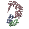
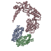
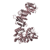
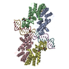
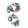
| Title | Membrane bound pleurotolysin prepore (TMH2 helix lock) trapped with engineered disulphide cross-link | ||||||
 Components Components |
| ||||||
 Keywords Keywords | TRANSPORT PROTEIN / MACPF/CDC SUPERFAMILY / PORE-FORMING PROTEINS | ||||||
| Function / homology |  Function and homology information Function and homology information | ||||||
| Biological species |  PLEUROTUS OSTREATUS (oyster mushroom) PLEUROTUS OSTREATUS (oyster mushroom) | ||||||
| Method | ELECTRON MICROSCOPY / single particle reconstruction / cryo EM / Resolution: 17 Å | ||||||
| Model type details | CA ATOMS ONLY, CHAIN A, B, C | ||||||
 Authors Authors | Lukoyanova, N. / Kondos, S.C. / Farabella, I. / Law, R.H.P. / Reboul, C.F. / Caradoc-Davies, T.T. / Spicer, B.A. / Kleifeld, O. / Perugini, M. / Ekkel, S. ...Lukoyanova, N. / Kondos, S.C. / Farabella, I. / Law, R.H.P. / Reboul, C.F. / Caradoc-Davies, T.T. / Spicer, B.A. / Kleifeld, O. / Perugini, M. / Ekkel, S. / Hatfaludi, T. / Oliver, K. / Hotze, E.M. / Tweten, R.K. / Whisstock, J.C. / Topf, M. / Dunstone, M.A. / Saibil, H.R. | ||||||
 Citation Citation | Journal: PLoS Biol / Year: 2015 Title: Conformational changes during pore formation by the perforin-related protein pleurotolysin. Authors: Natalya Lukoyanova / Stephanie C Kondos / Irene Farabella / Ruby H P Law / Cyril F Reboul / Tom T Caradoc-Davies / Bradley A Spicer / Oded Kleifeld / Daouda A K Traore / Susan M Ekkel / Ilia ...Authors: Natalya Lukoyanova / Stephanie C Kondos / Irene Farabella / Ruby H P Law / Cyril F Reboul / Tom T Caradoc-Davies / Bradley A Spicer / Oded Kleifeld / Daouda A K Traore / Susan M Ekkel / Ilia Voskoboinik / Joseph A Trapani / Tamas Hatfaludi / Katherine Oliver / Eileen M Hotze / Rodney K Tweten / James C Whisstock / Maya Topf / Helen R Saibil / Michelle A Dunstone /    Abstract: Membrane attack complex/perforin-like (MACPF) proteins comprise the largest superfamily of pore-forming proteins, playing crucial roles in immunity and pathogenesis. Soluble monomers assemble into ...Membrane attack complex/perforin-like (MACPF) proteins comprise the largest superfamily of pore-forming proteins, playing crucial roles in immunity and pathogenesis. Soluble monomers assemble into large transmembrane pores via conformational transitions that remain to be structurally and mechanistically characterised. Here we present an 11 Å resolution cryo-electron microscopy (cryo-EM) structure of the two-part, fungal toxin Pleurotolysin (Ply), together with crystal structures of both components (the lipid binding PlyA protein and the pore-forming MACPF component PlyB). These data reveal a 13-fold pore 80 Å in diameter and 100 Å in height, with each subunit comprised of a PlyB molecule atop a membrane bound dimer of PlyA. The resolution of the EM map, together with biophysical and computational experiments, allowed confident assignment of subdomains in a MACPF pore assembly. The major conformational changes in PlyB are a ∼70° opening of the bent and distorted central β-sheet of the MACPF domain, accompanied by extrusion and refolding of two α-helical regions into transmembrane β-hairpins (TMH1 and TMH2). We determined the structures of three different disulphide bond-trapped prepore intermediates. Analysis of these data by molecular modelling and flexible fitting allows us to generate a potential trajectory of β-sheet unbending. The results suggest that MACPF conformational change is triggered through disruption of the interface between a conserved helix-turn-helix motif and the top of TMH2. Following their release we propose that the transmembrane regions assemble into β-hairpins via top down zippering of backbone hydrogen bonds to form the membrane-inserted β-barrel. The intermediate structures of the MACPF domain during refolding into the β-barrel pore establish a structural paradigm for the transition from soluble monomer to pore, which may be conserved across the whole superfamily. The TMH2 region is critical for the release of both TMH clusters, suggesting why this region is targeted by endogenous inhibitors of MACPF function. | ||||||
| History |
|
- Structure visualization
| Movie |
Movie viewer |
|---|---|
| Structure viewer | Molecule: MolmilJmol/JSmol |
- Downloads & links
-Download
| PDBx/mmCIF format | 4v3m.cif.gz | 338.7 KB | Display | PDBx/mmCIF format |
|---|---|---|---|---|
| PDB format | pdb4v3m.ent.gz | 270.9 KB | Display | PDB format |
| PDBx/mmJSON format | 4v3m.json.gz | Tree view | PDBx/mmJSON format | |
| Others |  Other downloads Other downloads |
-Validation report
| Arichive directory | https://data.pdbj.org/pub/pdb/validation_reports/v3/4v3mftp://data.pdbj.org/pub/pdb/validation_reports/v3/4v3m | HTTPS FTP |
|---|
-Related structure data
| Related structure data | 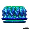 2795MC 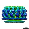 2793C  2794C  2796C 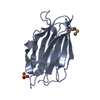 4oebC  4oejC  4ov8C 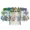 4v2tC 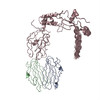 4v3aC 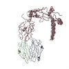 4v3nC C: citing same article ( M: map data used to model this data |
|---|---|
| Similar structure data |


-Links
 PDBj
PDBj- Assembly
| Deposited unit | 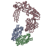
|
|---|---|
| 1 |
|
| Number of models | 20 |
-Components
| #1: Protein | Mass: 14852.545 Da / Num. of mol.: 2 Source method: isolated from a genetically manipulated source Source: (gene. exp.) PLEUROTUS OSTREATUS (oyster mushroom) / Plasmid: PET3A / Production host:  #2: Protein | | Mass: 57160.996 Da / Num. of mol.: 1 Source method: isolated from a genetically manipulated source Source: (gene. exp.) PLEUROTUS OSTREATUS (oyster mushroom) / Plasmid: PUC57, PET3A / Production host: |
|---|
-Experimental details
-Experiment
| Experiment | Method: ELECTRON MICROSCOPY |
|---|---|
| EM experiment | Aggregation state: PARTICLE / 3D reconstruction method: single particle reconstruction |
- Sample preparation
| Component | Name: PLEUROTOLYSIN PREPORE ON LIPOSOMES (TMH2 HELIX LOCK) TRAPPED WITH ENGINEERED DISULPHIDE Type: COMPLEX |
|---|---|
| Buffer solution | Name: 50 MM NACL, 20 MM HEPES / pH: 7.4 / Details: 50 MM NACL, 20 MM HEPES |
| Specimen | Conc.: 0.02 mg/ml / Embedding applied: NO / Shadowing applied: NO / Staining applied: NO / Vitrification applied: YES |
| Specimen support | Details: HOLEY CARBON |
| Vitrification | Instrument: FEI VITROBOT MARK III / Cryogen name: ETHANE Details: VITRIFICATION 1 -- CRYOGEN- ETHANE, HUMIDITY- 80,INSTRUMENT- FEI VITROBOT MARK III, METHOD- PLEUROTOLYSIN A WAS FIRST ADDED TO SPHINGOMYELIN-CHOLESTEROL LIPOSOMES AT A MOLAR RATIO OF 1 TO ...Details: VITRIFICATION 1 -- CRYOGEN- ETHANE, HUMIDITY- 80,INSTRUMENT- FEI VITROBOT MARK III, METHOD- PLEUROTOLYSIN A WAS FIRST ADDED TO SPHINGOMYELIN-CHOLESTEROL LIPOSOMES AT A MOLAR RATIO OF 1 TO 2000 ROTEIN TO LIPID IN THE ABOVE BUFFER. AFTER 5 MIN INCUBATION AT ROOM TEMPERATURE,PLEUROTOLYSIN B WAS ADDED TO THE MIXTURE AT A MOLAR RATIO OF 1 TO 2 TO PLEUROTOLYSIN A. THE MIXTURE WAS INCUBATED AT 40 C OR ROOM TEMPERATURE FOR 30 MIN AFTER WHICH 3.5 UL WERE PLACED ON NEGATIVELY GLOW DISCHARGED LACEY GRIDS AND VITRIFIED IN LIQUID ETHANE USING A VITROBOT. BLOTTING WAS CARRIED OUT AT 36 C AND 80 PERCENT HUMIDITY. |
- Electron microscopy imaging
| Experimental equipment |  Model: Tecnai Polara / Image courtesy: FEI Company |
|---|---|
| Microscopy | Model: FEI POLARA 300 / Date: Jan 30, 2013 |
| Electron gun | Electron source:  FIELD EMISSION GUN / Accelerating voltage: 300 kV / Illumination mode: FLOOD BEAM FIELD EMISSION GUN / Accelerating voltage: 300 kV / Illumination mode: FLOOD BEAM |
| Electron lens | Mode: BRIGHT FIELD / Nominal magnification: 59000 X / Calibrated magnification: 76148 X / Nominal defocus max: 3700 nm / Nominal defocus min: 700 nm / Cs: 2.3 mm |
| Specimen holder | Temperature: 94 K |
| Image recording | Electron dose: 25 e/Å2 / Film or detector model: GATAN ULTRASCAN 4000 (4k x 4k) |
| Image scans | Num. digital images: 154 |
| Radiation wavelength | Relative weight: 1 |
- Processing
| EM software |
| |||||||||||||||||||||
|---|---|---|---|---|---|---|---|---|---|---|---|---|---|---|---|---|---|---|---|---|---|---|
| CTF correction | Details: ESTIMATED WITH CTFFIND3, THEN PHASES FLIPPED FOR EACH PARTICLE | |||||||||||||||||||||
| Symmetry | Point symmetry: C13 (13 fold cyclic) | |||||||||||||||||||||
| 3D reconstruction | Method: ANGULAR RECONSTITUTION AND PROJECTION MATCHING / Resolution: 17 Å / Num. of particles: 722 / Nominal pixel size: 1.94 Å / Actual pixel size: 2 Å Details: UBMISSION BASED ON EXPERIMENTAL DATA FROM EMDB EMD-2795. (DEPOSITION ID: 12858). Symmetry type: POINT | |||||||||||||||||||||
| Atomic model building | Protocol: RIGID BODY FIT / Space: REAL / Target criteria: Cross-correlation coefficient / Details: METHOD--RIGID BODY | |||||||||||||||||||||
| Atomic model building | PDB-ID: 4OEB Accession code: 4OEB / Source name: PDB / Type: experimental model | |||||||||||||||||||||
| Refinement | Highest resolution: 17 Å | |||||||||||||||||||||
| Refinement step | Cycle: LAST / Highest resolution: 17 Å
|
