 Movie
Movie Controller


+ Open data

- Basic information
| Entry | Database: PDB / ID: 4pbg | ||||||
|---|---|---|---|---|---|---|---|
| Title | 6-PHOSPHO-BETA-GALACTOSIDASE FORM-CST | ||||||
 Components Components | 6-PHOSPHO-BETA-D-GALACTOSIDASE | ||||||
 Keywords Keywords | HYDROLASE / GLYCOSYL HYDROLASE | ||||||
| Function / homology |  Function and homology information Function and homology information6-phospho-beta-galactosidase / 6-phospho-beta-galactosidase activity / : / beta-glucosidase activity / cytosol Similarity search - Function | ||||||
| Biological species |  Lactococcus lactis (lactic acid bacteria) Lactococcus lactis (lactic acid bacteria) | ||||||
| Method |  X-RAY DIFFRACTION / MOLECULAR REPLACEMENT / Resolution: 2.5 Å X-RAY DIFFRACTION / MOLECULAR REPLACEMENT / Resolution: 2.5 Å | ||||||
 Authors Authors | Wiesmann, C. / Schulz, G.E. | ||||||
 Citation Citation | Journal: J.Mol.Biol. / Year: 1997 Title: Crystal structures and mechanism of 6-phospho-beta-galactosidase from Lactococcus lactis. Authors: Wiesmann, C. / Hengstenberg, W. / Schulz, G.E. #1: Journal: Eur.J.Biochem. / Year: 1995Title: Identification of the Active-Site Nucleophile in 6-Phospho-Beta-Galactosidase from Staphylococcus Aureus by Labelling with Synthetic Inhibitors Authors: Staedtler, P. / Hoenig, S. / Frank, R. / Withers, S.G. / Hengstenberg, W. #2: Journal: Structure / Year: 1995Title: The Three-Dimensional Structure of 6-Phospho-Beta-Galactosidase from Lactococcus Lactis Authors: Wiesmann, C. / Beste, G. / Hengstenberg, W. / Schulz, G.E. #3: Journal: Protein Eng. / Year: 1993Title: 6-Phospho-Beta-Galactosidases of Gram-Positive and 6-Phospho-Beta-Glucosidase B of Gram-Negative Bacteria: Comparison of Structure and Function by Kinetic and Immunological Methods and ...Title: 6-Phospho-Beta-Galactosidases of Gram-Positive and 6-Phospho-Beta-Glucosidase B of Gram-Negative Bacteria: Comparison of Structure and Function by Kinetic and Immunological Methods and Mutagenesis of the Lacg Gene of Staphylococcus Aureus Authors: Witt, E. / Frank, R. / Hengstenberg, W. | ||||||
| History |
|
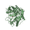
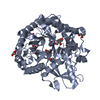
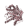
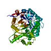
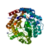
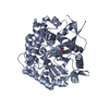
- Structure visualization
| Structure viewer | Molecule: MolmilJmol/JSmol |
|---|
- Downloads & links
-Download
| PDBx/mmCIF format | 4pbg.cif.gz | 203.2 KB | Display | PDBx/mmCIF format |
|---|---|---|---|---|
| PDB format | pdb4pbg.ent.gz | 163.7 KB | Display | PDB format |
| PDBx/mmJSON format | 4pbg.json.gz | Tree view | PDBx/mmJSON format | |
| Others |  Other downloads Other downloads |
-Validation report
| Arichive directory | https://data.pdbj.org/pub/pdb/validation_reports/pb/4pbgftp://data.pdbj.org/pub/pdb/validation_reports/pb/4pbg | HTTPS FTP |
|---|
-Related structure data
| Related structure data |  2pbgC 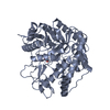 3pbgC  1pbgS S: Starting model for refinement C: citing same article ( |
|---|---|
| Similar structure data |


-Links
 PDBj
PDBj- Assembly
| Deposited unit | 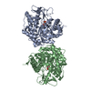
| ||||||||
|---|---|---|---|---|---|---|---|---|---|
| 1 | 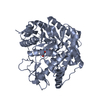
| ||||||||
| 2 | 
| ||||||||
| Unit cell |
| ||||||||
| Noncrystallographic symmetry (NCS) | NCS oper: (Code: given Matrix: (0.99987, 0.01597, 0.00069), Vector: |
-Components
| #1: Protein | Mass: 54113.141 Da / Num. of mol.: 2 / Mutation: G375C Source method: isolated from a genetically manipulated source Source: (gene. exp.) Lactococcus lactis (lactic acid bacteria)Strain: SUBSP. LACTIS 712 / Gene: LACG / Plasmid: PNZ316 / Gene (production host): LACG / Production host: #2: Sugar | 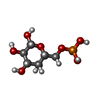  Type: D-saccharide, beta linking / Mass: 260.136 Da / Num. of mol.: 2 Type: D-saccharide, beta linking / Mass: 260.136 Da / Num. of mol.: 2Source method: isolated from a genetically manipulated source Formula: C6H13O9P #3: Water | ChemComp-HOH / |  Mass: 18.015 Da / Num. of mol.: 359 / Source method: isolated from a natural source / Formula: H2O Mass: 18.015 Da / Num. of mol.: 359 / Source method: isolated from a natural source / Formula: H2OCompound details | THE SECONDARY STRUCTURES HAVE BEEN ASSIGNED BY THE PROGRAM DSSP. THERE ARE SOME DISCREPANCIES WITH ...THE SECONDARY STRUCTURES | |
|---|
-Experimental details
-Experiment
| Experiment | Method: X-RAY DIFFRACTION / Number of used crystals: 1 |
|---|
- Sample preparation
| Crystal | Density Matthews: 2.7 Å3/Da / Density % sol: 55 % | ||||||||||||||||||||||||||||||||||||||||||||||||||||||
|---|---|---|---|---|---|---|---|---|---|---|---|---|---|---|---|---|---|---|---|---|---|---|---|---|---|---|---|---|---|---|---|---|---|---|---|---|---|---|---|---|---|---|---|---|---|---|---|---|---|---|---|---|---|---|---|
| Crystal grow | pH: 7.5 / Details: PEG-4000, PH 7.5 | ||||||||||||||||||||||||||||||||||||||||||||||||||||||
| Crystal grow | *PLUS Method: vapor diffusion, hanging drop | ||||||||||||||||||||||||||||||||||||||||||||||||||||||
| Components of the solutions | *PLUS
|
-Data collection
| Diffraction | Mean temperature: 293 K |
|---|---|
| Diffraction source | Source: ROTATING ANODE / Type: RIGAKU RUH2R / Wavelength: 1.5418 |
| Detector | Type: SIEMENS / Detector: AREA DETECTOR / Date: Jan 1, 1995 |
| Radiation | Monochromator: GRAPHITE(002) / Monochromatic (M) / Laue (L): M / Scattering type: x-ray |
| Radiation wavelength | Wavelength: 1.5418 Å / Relative weight: 1 |
| Reflection | Resolution: 2.5→1000 Å / Num. obs: 35989 / % possible obs: 84 % / Observed criterion σ(I): 0 / Redundancy: 4.1 % / Rsym value: 0.09 |
| Reflection | *PLUS Lowest resolution: 10 Å / Num. obs: 32734 / Rmerge(I) obs: 0.09 |
- Processing
| Software |
| ||||||||||||||||||||||||||||||||||||||||||||||||||||||||||||
|---|---|---|---|---|---|---|---|---|---|---|---|---|---|---|---|---|---|---|---|---|---|---|---|---|---|---|---|---|---|---|---|---|---|---|---|---|---|---|---|---|---|---|---|---|---|---|---|---|---|---|---|---|---|---|---|---|---|---|---|---|---|
| Refinement | Method to determine structure: MOLECULAR REPLACEMENT Starting model: PDB ENTRY 1PBG Resolution: 2.5→10 Å / Data cutoff high absF: 100000 / Data cutoff low absF: 0 / σ(F): 0
| ||||||||||||||||||||||||||||||||||||||||||||||||||||||||||||
| Displacement parameters | Biso mean: 14 Å2 | ||||||||||||||||||||||||||||||||||||||||||||||||||||||||||||
| Refine analyze | Luzzati coordinate error obs: 0.28 Å / Luzzati sigma a obs: 0.38 Å | ||||||||||||||||||||||||||||||||||||||||||||||||||||||||||||
| Refinement step | Cycle: LAST / Resolution: 2.5→10 Å
| ||||||||||||||||||||||||||||||||||||||||||||||||||||||||||||
| Refine LS restraints |
| ||||||||||||||||||||||||||||||||||||||||||||||||||||||||||||
| Software | *PLUS Name: X-PLOR / Classification: refinement | ||||||||||||||||||||||||||||||||||||||||||||||||||||||||||||
| Refinement | *PLUS | ||||||||||||||||||||||||||||||||||||||||||||||||||||||||||||
| Solvent computation | *PLUS | ||||||||||||||||||||||||||||||||||||||||||||||||||||||||||||
| Displacement parameters | *PLUS | ||||||||||||||||||||||||||||||||||||||||||||||||||||||||||||
| Refine LS restraints | *PLUS
|
