 Movie
Movie Controller

[English] 日本語
 Yorodumi
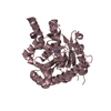
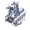
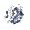
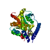
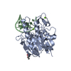
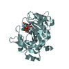
- PDB-4n5i: Crystal Structure of a C8-C4 Sn3 Inhibited Esterase B from Lactob... -
+ Open data

- Basic information
| Entry | Database: PDB / ID: 4n5i | ||||||
|---|---|---|---|---|---|---|---|
| Title | Crystal Structure of a C8-C4 Sn3 Inhibited Esterase B from Lactobacillus Rhamnosis | ||||||
 Components Components | Esterase/lipase | ||||||
 Keywords Keywords | HYDROLASE / alpha/beta Hydrolase Fold / Esterase/Lipase Transferase / Triacylglycerase / Hydrolysis | ||||||
| Function / homology | Alpha/Beta hydrolase fold, catalytic domain / Rossmann fold / 3-Layer(aba) Sandwich / Alpha Beta / (2R)-2,3-dibutoxypropyl (R)-octylphosphinate / ACETATE ION / :  Function and homology information Function and homology information | ||||||
| Biological species |  Lactobacillus rhamnosus (bacteria) Lactobacillus rhamnosus (bacteria) | ||||||
| Method |  X-RAY DIFFRACTION / MOLECULAR REPLACEMENT / Resolution: 2 Å X-RAY DIFFRACTION / MOLECULAR REPLACEMENT / Resolution: 2 Å | ||||||
 Authors Authors | Colbert, D.A. / Bennett, M.D. / Lun, D.J. / Loo, T.S. / Anderson, B.F. / Norris, G.E. | ||||||
 Citation Citation | Journal: TO BE PUBLISHED Title: Crystal Structure of a C8-C4 Sn3 Inhibited Esterase B from Lactobacillus Rhamnosis Authors: Colbert, D.A. / D Bennett, M. / J Lun, D. / S Loo, T. / F Anderson, B. / E Norris, G. | ||||||
| History |
|
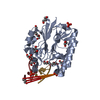
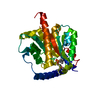
- Structure visualization
| Structure viewer | Molecule: MolmilJmol/JSmol |
|---|
- Downloads & links
-Download
| PDBx/mmCIF format | 4n5i.cif.gz | 84 KB | Display | PDBx/mmCIF format |
|---|---|---|---|---|
| PDB format | pdb4n5i.ent.gz | 61.9 KB | Display | PDB format |
| PDBx/mmJSON format | 4n5i.json.gz | Tree view | PDBx/mmJSON format | |
| Others |  Other downloads Other downloads |
-Validation report
| Arichive directory | https://data.pdbj.org/pub/pdb/validation_reports/n5/4n5iftp://data.pdbj.org/pub/pdb/validation_reports/n5/4n5i | HTTPS FTP |
|---|
-Related structure data
| Related structure data | 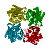 4n5hS S: Starting model for refinement |
|---|---|
| Similar structure data |

-Links
 PDBj
PDBj- Assembly
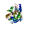
| Deposited unit | 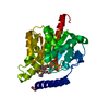
| ||||||||
|---|---|---|---|---|---|---|---|---|---|
| 1 |
| ||||||||
| Unit cell |
|
-Components
| #1: Protein | Mass: 35794.027 Da / Num. of mol.: 1 / Mutation: V210A Source method: isolated from a genetically manipulated source Source: (gene. exp.) Lactobacillus rhamnosus (bacteria) / Strain: Lc 705 / Gene: LC705_02872, LRH_10360 / Plasmid: pGEX_6P3_EstB / Production host: | ||
|---|---|---|---|
| #2: Chemical | ChemComp-2HD / (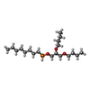  Mass: 364.500 Da / Num. of mol.: 1 / Source method: obtained synthetically / Formula: C19H41O4P Mass: 364.500 Da / Num. of mol.: 1 / Source method: obtained synthetically / Formula: C19H41O4P | ||
| #3: Chemical |   Mass: 59.044 Da / Num. of mol.: 2 / Source method: obtained synthetically / Formula: C2H3O2 Mass: 59.044 Da / Num. of mol.: 2 / Source method: obtained synthetically / Formula: C2H3O2#4: Water | ChemComp-HOH / |  Mass: 18.015 Da / Num. of mol.: 297 / Source method: isolated from a natural source / Formula: H2O Mass: 18.015 Da / Num. of mol.: 297 / Source method: isolated from a natural source / Formula: H2O |
-Experimental details
-Experiment
| Experiment | Method: X-RAY DIFFRACTION / Number of used crystals: 1 |
|---|
- Sample preparation
| Crystal | Density Matthews: 2.5 Å3/Da / Density % sol: 50.8 % |
|---|---|
| Crystal grow | Temperature: 293 K / Method: vapor diffusion, hanging drop / pH: 8.5 Details: 1.0 M ammonium phosphate, 0.1 M TrisHCl, pH 7.2-8.5 , VAPOR DIFFUSION, HANGING DROP, temperature 293K |
-Data collection
| Diffraction | Mean temperature: 110 K |
|---|---|
| Diffraction source | Source: ROTATING ANODE / Type: RIGAKU MICROMAX-002 / Wavelength: 1.5418 Å |
| Detector | Type: RIGAKU RAXIS IV++ / Detector: IMAGE PLATE / Date: Jun 28, 2007 / Details: Capillary focusing optics and monochromator |
| Radiation | Monochromator: capillary focusing optics and monochromator / Protocol: SINGLE WAVELENGTH / Monochromatic (M) / Laue (L): M / Scattering type: x-ray |
| Radiation wavelength | Wavelength: 1.5418 Å / Relative weight: 1 |
| Reflection | Resolution: 2→27.1 Å / Num. all: 23508 / Num. obs: 23508 / % possible obs: 97 % / Observed criterion σ(I): 3 / Redundancy: 4.17 % / Biso Wilson estimate: 21.6 Å2 / Rmerge(I) obs: 0.116 / Net I/σ(I): 6.6 |
| Reflection shell | Resolution: 2→2.07 Å / Redundancy: 4.14 % / Rmerge(I) obs: 0.4 / Mean I/σ(I) obs: 2.4 / Num. unique all: 2269 / % possible all: 95.9 |
- Processing
| Software |
| ||||||||||||||||||||||||||||||||||||||||||||||||||||||||||||
|---|---|---|---|---|---|---|---|---|---|---|---|---|---|---|---|---|---|---|---|---|---|---|---|---|---|---|---|---|---|---|---|---|---|---|---|---|---|---|---|---|---|---|---|---|---|---|---|---|---|---|---|---|---|---|---|---|---|---|---|---|---|
| Refinement | Method to determine structure: MOLECULAR REPLACEMENT Starting model: PDB entry 4N5H Resolution: 2→27.1 Å / Cor.coef. Fo:Fc: 0.962 / Cor.coef. Fo:Fc free: 0.937 / SU B: 3.665 / SU ML: 0.102 / Isotropic thermal model: Overall / Cross valid method: THROUGHOUT / ESU R: 0.169 / ESU R Free: 0.157 / Stereochemistry target values: MAXIMUM LIKELIHOOD / Details: HYDROGENS HAVE BEEN ADDED IN THE RIDING POSITIONS
| ||||||||||||||||||||||||||||||||||||||||||||||||||||||||||||
| Solvent computation | Ion probe radii: 0.8 Å / Shrinkage radii: 0.8 Å / VDW probe radii: 1.2 Å / Solvent model: MASK | ||||||||||||||||||||||||||||||||||||||||||||||||||||||||||||
| Displacement parameters | Biso mean: 22.402 Å2
| ||||||||||||||||||||||||||||||||||||||||||||||||||||||||||||
| Refinement step | Cycle: LAST / Resolution: 2→27.1 Å
| ||||||||||||||||||||||||||||||||||||||||||||||||||||||||||||
| Refine LS restraints |
| ||||||||||||||||||||||||||||||||||||||||||||||||||||||||||||
| LS refinement shell | Resolution: 2→2.052 Å / Total num. of bins used: 20
|
