 Movie
Movie Controller

[English] 日本語
 Yorodumi
- PDB-4i9n: Crystal structure of rabbit LDHA in complex with AP28161 and AP28122 -
+ Open data

- Basic information
| Entry | Database: PDB / ID: 4i9n | ||||||
|---|---|---|---|---|---|---|---|
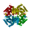
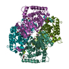
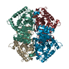
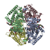
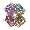
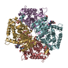
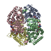
| Title | Crystal structure of rabbit LDHA in complex with AP28161 and AP28122 | ||||||
 Components Components | L-lactate dehydrogenase A chain | ||||||
 Keywords Keywords | OXIDOREDUCTASE/OXIDOREDUCTASE INHIBITOR / cancer / fragment / inhibitor / OXIDOREDUCTASE-OXIDOREDUCTASE INHIBITOR complex | ||||||
| Function / homology |  Function and homology information Function and homology informationL-lactate dehydrogenase / L-lactate dehydrogenase (NAD+) activity / lactate metabolic process / cytoplasm Similarity search - Function | ||||||
| Biological species |  | ||||||
| Method |  X-RAY DIFFRACTION / MOLECULAR REPLACEMENT / Resolution: 2.35 Å X-RAY DIFFRACTION / MOLECULAR REPLACEMENT / Resolution: 2.35 Å | ||||||
 Authors Authors | Zhou, T. / Kohlmann, A. / Stephan, Z.G. / Li, F. / Commodore, L. / Greenfield, M.T. / Shakespeare, W.C. / Zhu, X. / Dalgarno, D.C. | ||||||
 Citation Citation | Journal: J.Med.Chem. / Year: 2013 Title: Fragment growing and linking lead to novel nanomolar lactate dehydrogenase inhibitors. Authors: Kohlmann, A. / Zech, S.G. / Li, F. / Zhou, T. / Squillace, R.M. / Commodore, L. / Greenfield, M.T. / Lu, X. / Miller, D.P. / Huang, W.S. / Qi, J. / Thomas, R.M. / Wang, Y. / Zhang, S. / ...Authors: Kohlmann, A. / Zech, S.G. / Li, F. / Zhou, T. / Squillace, R.M. / Commodore, L. / Greenfield, M.T. / Lu, X. / Miller, D.P. / Huang, W.S. / Qi, J. / Thomas, R.M. / Wang, Y. / Zhang, S. / Dodd, R. / Liu, S. / Xu, R. / Xu, Y. / Miret, J.J. / Rivera, V. / Clackson, T. / Shakespeare, W.C. / Zhu, X. / Dalgarno, D.C. | ||||||
| History |
|
- Structure visualization
| Structure viewer | Molecule: MolmilJmol/JSmol |
|---|
- Downloads & links
-Download
| PDBx/mmCIF format | 4i9n.cif.gz | 504.1 KB | Display | PDBx/mmCIF format |
|---|---|---|---|---|
| PDB format | pdb4i9n.ent.gz | 420.7 KB | Display | PDB format |
| PDBx/mmJSON format | 4i9n.json.gz | Tree view | PDBx/mmJSON format | |
| Others |  Other downloads Other downloads |
-Validation report
| Arichive directory | https://data.pdbj.org/pub/pdb/validation_reports/i9/4i9nftp://data.pdbj.org/pub/pdb/validation_reports/i9/4i9n | HTTPS FTP |
|---|
-Related structure data
| Related structure data |  4i8xC  4i9hC 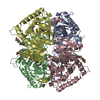 4i9uC  1i10S C: citing same article ( S: Starting model for refinement |
|---|---|
| Similar structure data |


-Links
 PDBj
PDBj


- Assembly
| Deposited unit | 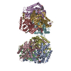
| ||||||||
|---|---|---|---|---|---|---|---|---|---|
| 1 | 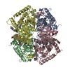
| ||||||||
| 2 | 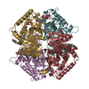
| ||||||||
| Unit cell |
|
-Components
| #1: Protein | Mass: 36481.375 Da / Num. of mol.: 8 / Source method: isolated from a natural source / Source: (natural) #2: Chemical | ChemComp-1E6 /   Mass: 442.871 Da / Num. of mol.: 8 / Source method: obtained synthetically / Formula: C18H19ClN2O7S Mass: 442.871 Da / Num. of mol.: 8 / Source method: obtained synthetically / Formula: C18H19ClN2O7S#3: Chemical | ChemComp-1E5 /   Mass: 291.231 Da / Num. of mol.: 8 / Source method: obtained synthetically / Formula: C14H10FNO5 Mass: 291.231 Da / Num. of mol.: 8 / Source method: obtained synthetically / Formula: C14H10FNO5#4: Water | ChemComp-HOH / |  Mass: 18.015 Da / Num. of mol.: 313 / Source method: isolated from a natural source / Formula: H2O Mass: 18.015 Da / Num. of mol.: 313 / Source method: isolated from a natural source / Formula: H2O |
|---|
-Experimental details
-Experiment
| Experiment | Method: X-RAY DIFFRACTION / Number of used crystals: 1 |
|---|
- Sample preparation
| Crystal | Density Matthews: 2.79 Å3/Da / Density % sol: 55.88 % |
|---|---|
| Crystal grow | Temperature: 277 K / Method: vapor diffusion, hanging drop / pH: 8 Details: 0.1 M Tris, pH 8.0, 30% PEG550 MME, VAPOR DIFFUSION, HANGING DROP, temperature 277K |
-Data collection
| Diffraction | Mean temperature: 100 K |
|---|---|
| Diffraction source | Source: ROTATING ANODE / Type: RIGAKU / Wavelength: 1.54 Å |
| Detector | Type: RIGAKU RAXIS IV++ / Detector: IMAGE PLATE / Date: Feb 8, 2010 |
| Radiation | Protocol: SINGLE WAVELENGTH / Monochromatic (M) / Laue (L): M / Scattering type: x-ray |
| Radiation wavelength | Wavelength: 1.54 Å / Relative weight: 1 |
| Reflection | Resolution: 2.35→50 Å / Num. all: 141773 / Num. obs: 133852 / % possible obs: 94.4 % / Observed criterion σ(F): 2 / Observed criterion σ(I): 2 |
| Reflection shell | Highest resolution: 2.35 Å |
- Processing
| Software |
| ||||||||||||||||||||||||||||
|---|---|---|---|---|---|---|---|---|---|---|---|---|---|---|---|---|---|---|---|---|---|---|---|---|---|---|---|---|---|
| Refinement | Method to determine structure: MOLECULAR REPLACEMENT Starting model: PDB ENTRY 1I10 Resolution: 2.35→50 Å / Occupancy max: 1 / Occupancy min: 0.5 / σ(F): 0 / Stereochemistry target values: Engh & Huber
| ||||||||||||||||||||||||||||
| Displacement parameters | Biso max: 96.32 Å2 / Biso mean: 30.9829 Å2 / Biso min: 3.65 Å2
| ||||||||||||||||||||||||||||
| Refinement step | Cycle: LAST / Resolution: 2.35→50 Å
| ||||||||||||||||||||||||||||
| Refine LS restraints |
| ||||||||||||||||||||||||||||
| Xplor file |
|
