 Movie
Movie Controller

[English] 日本語
 Yorodumi
- PDB-6ldh: REFINED CRYSTAL STRUCTURE OF DOGFISH M4 APO-LACTATE DEHYDROGENASE -
+ Open data

- Basic information
| Entry | Database: PDB / ID: 6ldh | |||||||||
|---|---|---|---|---|---|---|---|---|---|---|
| Title | REFINED CRYSTAL STRUCTURE OF DOGFISH M4 APO-LACTATE DEHYDROGENASE | |||||||||
 Components Components | M4 APO-LACTATE DEHYDROGENASE | |||||||||
 Keywords Keywords | OXIDOREDUCTASE(CHOH(D)-NAD(A)) | |||||||||
| Function / homology |  Function and homology information Function and homology informationL-lactate dehydrogenase / L-lactate dehydrogenase (NAD+) activity / lactate metabolic process / cytoplasm Similarity search - Function | |||||||||
| Biological species |  Squalus acanthias (spiny dogfish) Squalus acanthias (spiny dogfish) | |||||||||
| Method |  X-RAY DIFFRACTION / Resolution: 2 Å X-RAY DIFFRACTION / Resolution: 2 Å | |||||||||
 Authors Authors | Abad-Zapatero, C. / Rossmann, M.G. | |||||||||
 Citation Citation | Journal: J.Mol.Biol. / Year: 1987 Title: Refined crystal structure of dogfish M4 apo-lactate dehydrogenase. Authors: Abad-Zapatero, C. / Griffith, J.P. / Sussman, J.L. / Rossmann, M.G. #1: Journal: J.Mol.Biol. / Year: 1976Title: A Comparison of the Structures of Apo Dogfish M4 Lactate Dehydrogenase and its Ternary Complexes Authors: White, J.L. / Hackert, M.L. / Buehner, M. / Adams, M.J. / Ford, G.C. / Lentzjunior, P.J. / Smiley, I.E. / Steindel, S.J. / Rossmann, M.G. #2: Journal: Proc.Natl.Acad.Sci.USA / Year: 1977Title: Structural Adaptations of Lactate Dehydrogenase Isozymes Authors: Eventoff, W. / Rossmann, M.G. / Taylor, S.S. / Torff, H.-J. / Meyer, H. / Keil, W. / Kiltz, H.-H. #3: Journal: J.Mol.Biol. / Year: 1975Title: A 5 Angstroms X-Ray Diffraction Study of Coenzyme-Deficient Lactate Dehydrogenase,Nad-Pyruvate Ternary Complex Authors: White, J. / Rossmann, M.G. / Ford, G.C. #4: Journal: Isozymes-Molecular Structure / Year: 1975Title: A Structural Comparison of Porcine B4 and Dogfish A4 Isozymes of Lactate Dehydrogenase Authors: Eventoff, W. / Hackert, M.L. / Steindel, S.J. / Rossmann, M.G. #5: Journal: The Enzymes,Third Edition / Year: 1975Title: Lactate Dehydrogenase Authors: Holbrook, J.J. / Liljas, A. / Steindel, S.J. / Rossmann, M.G. #6: Journal: Annu.Rev.Biochem. / Year: 1974Title: X-Ray Studies of Protein Interactions Authors: Liljas, A. / Rossmann, M.G. #7: Journal: Nature / Year: 1974Title: Chemical and Biological Evolution of a Nucleotide Binding Protein Authors: Rossmann, M.G. / Moras, D. / Olsen, K.W. #8: Journal: J.Mol.Biol. / Year: 1974Title: Recognition of Structural Domains in Globular Proteins Authors: Rossmann, M.G. / Liljas, A. #9: Journal: Biochem.Biophys.Res.Commun. / Year: 1973Title: Atomic Co-Ordinates for Dogfish M4 Apo-Lactate Dehydrogenase Authors: Adams, M.J. / Ford, G.C. / Liljas, A. / Rossmann, M.G. #10: Journal: Proc.Natl.Acad.Sci.USA / Year: 1973Title: Structure-Function Relationships in Lactate Dehydrogenase Authors: Adams, M.J. / Buehner, M. / Chandrasekhar, K. / Ford, G.C. / Hackert, M.L. / Liljas, A. / Rossmann, M.G. / Smiley, I.E. / Allison, W.S. / Everse, J. / Kaplan, N.O. / Taylor, S.S. #11: Journal: Proc.Natl.Acad.Sci.USA / Year: 1973Title: Amino-Acid Sequence of Dogfish M4 Lactate Dehydrogenase Authors: Taylor, S.S. / Oxley, S.S. / Allison, W.S. / Kaplan, N.O. #12: Journal: J.Mol.Biol. / Year: 1973Title: Molecular Symmetry Axes and Subunit Interfaces in Certain Dehydrogenases Authors: Rossmann, M.G. / Adams, M.J. / Buehner, M. / Ford, G.C. / Hackert, M.L. / Liljas, A. / Rao, S.T. / Banaszak, L.J. / Hill, E. / Tsernoglou, D. / Webb, L. #13: Journal: J.Mol.Biol. / Year: 1973Title: Functional Anion Binding Sites in Dogfish M4 Lactate Dehydrogenase Authors: Adams, M.J. / Liljas, A. / Rossmann, M.G. #14: Journal: J.Mol.Biol. / Year: 1973Title: Binding of Oxamate to the Apoenzyme of Dogfish M4 Lactate Dehydrogenase Authors: Mcphersonjunior, A. #15: Journal: J.Mol.Biol. / Year: 1973Title: Conformation of Coenzyme Fragments When Bound to Lactate Dehydrogenase Authors: Chandrasekhar, K. / Mcphersonjunior, A. / Adams, M.J. / Rossmann, M.G. #16: Journal: J.Mol.Biol. / Year: 1971Title: The 5 Angstroms Resolution Structure of an Abortive Ternary Complex of Lactate Dehydrogenase and its Comparison with the Apo-Enzyme Authors: Smiley, I.E. / Koekoek, R. / Adams, M.J. / Rossmann, M.G. #17: Journal: Nature / Year: 1970Title: Structure of Lactate Dehydrogenase at 2.8 Angstroms Resolution Authors: Adams, M.J. / Ford, G.C. / Koekoek, R. / Lentzjunior, P.J. / Mcphersonjunior, A. / Rossmann, M.G. / Smiley, I.E. / Schevitz, R.W. / Wonacott, A.J. #18: Journal: J.Mol.Biol. / Year: 1969Title: Low Resolution Study of Crystalline L-Lactate Dehydrogenase Authors: Adams, M.J. / Haas, D.J. / Jeffery, B.A. / Mcphersonjunior, A. / Mermall, H.L. / Rossmann, M.G. / Schevitz, R.W. / Wonacott, A.J. #19: Journal: Proc.Natl.Acad.Sci.USA / Year: 1967Title: The Crystal Structure of Lactic Dehydrogenase Authors: Rossmann, M.G. / Jeffery, B.A. / Main, P. / Warren, S. | |||||||||
| History |
| |||||||||
| Remark 700 | SHEET STRAND 2 OF SHEET S2 IS BIFURCATED. THIS IS REPRESENTED BY TWO SHEETS, S2A AND S2B BELOW, ...SHEET STRAND 2 OF SHEET S2 IS BIFURCATED. THIS IS REPRESENTED BY TWO SHEETS, S2A AND S2B BELOW, WHERE THE FIRST STRAND OF IS S2A IS IDENTICAL TO THE FIRST STRAND OF S2B. |
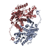
- Structure visualization
| Structure viewer | Molecule: MolmilJmol/JSmol |
|---|
- Downloads & links
-Download
| PDBx/mmCIF format | 6ldh.cif.gz | 85.6 KB | Display | PDBx/mmCIF format |
|---|---|---|---|---|
| PDB format | pdb6ldh.ent.gz | 63.8 KB | Display | PDB format |
| PDBx/mmJSON format | 6ldh.json.gz | Tree view | PDBx/mmJSON format | |
| Others |  Other downloads Other downloads |
-Validation report
| Arichive directory | https://data.pdbj.org/pub/pdb/validation_reports/ld/6ldhftp://data.pdbj.org/pub/pdb/validation_reports/ld/6ldh | HTTPS FTP |
|---|
-Related structure data

-Links
 PDBj
PDBj


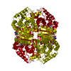
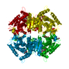
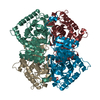
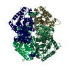
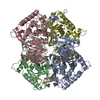
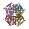
- Assembly
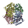
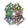
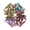
| Deposited unit | 
| ||||||||
|---|---|---|---|---|---|---|---|---|---|
| 1 | 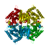
| ||||||||
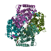
| Unit cell |
| ||||||||
| Atom site foot note | 1: RESIDUE PRO 139 IS A CIS PROLINE. | ||||||||
| Details | NON-CRYSTALLOGRAPHIC SYMMETRY ELEMENTS CORRESPONDING TO THE THREE ORTHOGONAL MOLECULAR SYMMETRY AXES ARE PRESENTED ON THE MTRIX RECORDS BELOW. |
-Components
| #1: Protein | Mass: 36353.297 Da / Num. of mol.: 1 Source method: isolated from a genetically manipulated source Source: (gene. exp.) Squalus acanthias (spiny dogfish) / References: UniProt: P00341, L-lactate dehydrogenase | ||||||
|---|---|---|---|---|---|---|---|
| #2: Chemical |   Mass: 96.063 Da / Num. of mol.: 2 / Source method: obtained synthetically / Formula: SO4 Mass: 96.063 Da / Num. of mol.: 2 / Source method: obtained synthetically / Formula: SO4#3: Water | ChemComp-HOH / |  Mass: 18.015 Da / Num. of mol.: 286 / Source method: isolated from a natural source / Formula: H2O Mass: 18.015 Da / Num. of mol.: 286 / Source method: isolated from a natural source / Formula: H2OHas protein modification | Y | Sequence details | THE RESIDUES IN THIS ENTRY ARE NUMBERED SEQUENTIALLY FROM 0 - 329. SEE THE PAPER CITED AS REFERENCE ...THE RESIDUES IN THIS ENTRY ARE NUMBERED SEQUENTIAL | |
-Experimental details
-Experiment
| Experiment | Method: X-RAY DIFFRACTION |
|---|
- Sample preparation
| Crystal | Density Matthews: 2.88 Å3/Da / Density % sol: 57.26 % |
|---|---|
| Crystal grow | *PLUS Method: vapor diffusion, hanging dropDetails: referred to 'Pesca, A. J.', (1967) J.Biol.Chem., 242, 2151-2167 |
| Components of the solutions | *PLUS Conc.: 30 %sat / Common name: ammonium sulfate |
-Data collection
| Radiation | Scattering type: x-ray |
|---|---|
| Radiation wavelength | Relative weight: 1 |
- Processing
| Software | Name: PROLSQ / Classification: refinement | ||||||||||||||||||||||||||||||||||||||||||||||||||||||||||||||||||||||||||||||||||||
|---|---|---|---|---|---|---|---|---|---|---|---|---|---|---|---|---|---|---|---|---|---|---|---|---|---|---|---|---|---|---|---|---|---|---|---|---|---|---|---|---|---|---|---|---|---|---|---|---|---|---|---|---|---|---|---|---|---|---|---|---|---|---|---|---|---|---|---|---|---|---|---|---|---|---|---|---|---|---|---|---|---|---|---|---|---|
| Refinement | Resolution: 2→8 Å / Rfactor obs: 0.202 Details: SPACE GROUP F 4 2 2 IS A NON-STANDARD REPRESENTATION OF THE GROUP I 4 2 2. IN THIS CASE THE AXES OF THE UNIT CELL ARE CONSIDERED TO BE LEFT-HANDED. USING CONVENTIONAL NOTATION THE EQUI- ...Details: SPACE GROUP F 4 2 2 IS A NON-STANDARD REPRESENTATION OF THE GROUP I 4 2 2. IN THIS CASE THE AXES OF THE UNIT CELL ARE CONSIDERED TO BE LEFT-HANDED. USING CONVENTIONAL NOTATION THE EQUI-POINTS OF THE F 4 2 2 CELL MAY BE EXPRESSED AS- ( 0,0,0 0,1/2,1/2 1/2,0,1/2 1/2,1/2,0 ) + X, Y, Z -X,-Y, Z -X, Y,-Z X,-Y,-Z Y, X,-Z -Y,-X,-Z -Y, X, Z Y,-X, Z . | ||||||||||||||||||||||||||||||||||||||||||||||||||||||||||||||||||||||||||||||||||||
| Refinement step | Cycle: LAST / Resolution: 2→8 Å
| ||||||||||||||||||||||||||||||||||||||||||||||||||||||||||||||||||||||||||||||||||||
| Refine LS restraints |
| ||||||||||||||||||||||||||||||||||||||||||||||||||||||||||||||||||||||||||||||||||||
| Refinement | *PLUS Highest resolution: 2 Å / Lowest resolution: 8 Å / Rfactor obs: 0.202 | ||||||||||||||||||||||||||||||||||||||||||||||||||||||||||||||||||||||||||||||||||||
| Solvent computation | *PLUS | ||||||||||||||||||||||||||||||||||||||||||||||||||||||||||||||||||||||||||||||||||||
| Displacement parameters | *PLUS |
