Movie
Movie Controller


+ Open data

- Basic information
| Entry | Database: EMDB / ID: EMD-4322 | |||||||||
|---|---|---|---|---|---|---|---|---|---|---|
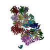
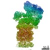
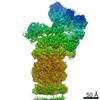
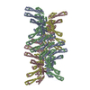
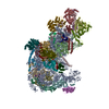
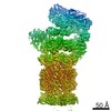
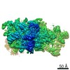
| Title | 26S proteasome, s4 state | |||||||||
 Map data Map data | None | |||||||||
 Sample Sample |
| |||||||||
 Keywords Keywords | 26S proteasome / AAA+ ATPase / hydrolase | |||||||||
| Function / homology |  Function and homology information Function and homology informationSAGA complex localization to transcription regulatory region / : / proteasome regulatory particle assembly / proteasome storage granule assembly / peroxisome fission / transcription export complex 2 / maintenance of DNA trinucleotide repeats / protein deneddylation / filamentous growth / COP9 signalosome ...SAGA complex localization to transcription regulatory region / : / proteasome regulatory particle assembly / proteasome storage granule assembly / peroxisome fission / transcription export complex 2 / maintenance of DNA trinucleotide repeats / protein deneddylation / filamentous growth / COP9 signalosome / mitochondrial fission / proteasome regulatory particle / proteasome-activating activity / proteasome regulatory particle, lid subcomplex / proteasome regulatory particle, base subcomplex / ER-Phagosome pathway / Antigen processing: Ub, ATP-independent proteasomal degradation / protein-containing complex localization / proteasome core complex assembly / Proteasome assembly / Cross-presentation of soluble exogenous antigens (endosomes) / TNFR2 non-canonical NF-kB pathway / K48-linked polyubiquitin modification-dependent protein binding / nuclear outer membrane-endoplasmic reticulum membrane network / nonfunctional rRNA decay / Regulation of PTEN stability and activity / CDK-mediated phosphorylation and removal of Cdc6 / metal-dependent deubiquitinase activity / FBXL7 down-regulates AURKA during mitotic entry and in early mitosis / KEAP1-NFE2L2 pathway / Neddylation / peptide catabolic process / Ubiquitin-Mediated Degradation of Phosphorylated Cdc25A / Orc1 removal from chromatin / MAPK6/MAPK4 signaling / proteasome binding / Antigen processing: Ubiquitination & Proteasome degradation / regulation of protein catabolic process / proteasome storage granule / proteasomal ubiquitin-independent protein catabolic process / positive regulation of RNA polymerase II transcription preinitiation complex assembly / Ub-specific processing proteases / protein deubiquitination / polyubiquitin modification-dependent protein binding / proteasome endopeptidase complex / proteasome core complex, beta-subunit complex / endopeptidase activator activity / threonine-type endopeptidase activity / proteasome core complex, alpha-subunit complex / mRNA export from nucleus / proteasome assembly / enzyme regulator activity / Neutrophil degranulation / ERAD pathway / protein folding chaperone / proteasome complex / nucleotide-excision repair / ubiquitin binding / positive regulation of transcription elongation by RNA polymerase II / double-strand break repair via homologous recombination / metallopeptidase activity / positive regulation of protein catabolic process / peroxisome / positive regulation of proteasomal ubiquitin-dependent protein catabolic process / endopeptidase activity / molecular adaptor activity / ubiquitin-dependent protein catabolic process / proteasome-mediated ubiquitin-dependent protein catabolic process / protein-macromolecule adaptor activity / cysteine-type deubiquitinase activity / ubiquitinyl hydrolase 1 / regulation of cell cycle / chromatin remodeling / protein domain specific binding / mRNA binding / ubiquitin protein ligase binding / endoplasmic reticulum membrane / structural molecule activity / endoplasmic reticulum / ATP hydrolysis activity / positive regulation of transcription by RNA polymerase II / mitochondrion / ATP binding / metal ion binding / identical protein binding / nucleus / cytoplasm / cytosol Similarity search - Function | |||||||||
| Biological species |  | |||||||||
| Method | single particle reconstruction / cryo EM / Resolution: 4.5 Å | |||||||||
 Authors Authors | Eisele MR / Reed RG | |||||||||
 Citation Citation | Journal: Cell Rep / Year: 2018 Title: Expanded Coverage of the 26S Proteasome Conformational Landscape Reveals Mechanisms of Peptidase Gating. Authors: Markus R Eisele / Randi G Reed / Till Rudack / Andreas Schweitzer / Florian Beck / Istvan Nagy / Günter Pfeifer / Jürgen M Plitzko / Wolfgang Baumeister / Robert J Tomko / Eri Sakata /   Abstract: The proteasome is the central protease for intracellular protein breakdown. Coordinated binding and hydrolysis of ATP by the six proteasomal ATPase subunits induces conformational changes that drive ...The proteasome is the central protease for intracellular protein breakdown. Coordinated binding and hydrolysis of ATP by the six proteasomal ATPase subunits induces conformational changes that drive the unfolding and translocation of substrates into the proteolytic 20S core particle for degradation. Here, we combine genetic and biochemical approaches with cryo-electron microscopy and integrative modeling to dissect the relationship between individual nucleotide binding events and proteasome conformational dynamics. We demonstrate unique impacts of ATP binding by individual ATPases on the proteasome conformational distribution and report two conformational states of the proteasome suggestive of a rotary ATP hydrolysis mechanism. These structures, coupled with functional analyses, reveal key roles for the ATPases Rpt1 and Rpt6 in gating substrate entry into the core particle. This deepened knowledge of proteasome conformational dynamics reveals key elements of intersubunit communication within the proteasome and clarifies the regulation of substrate entry into the proteolytic chamber. | |||||||||
| History |
|
- Structure visualization
| Movie |
Movie viewer |
|---|---|
| Structure viewer | EM map: SurfViewMolmilJmol/JSmol |
| Supplemental images |

- Downloads & links
-EMDB archive
| Map data | emd_4322.map.gz | 192.6 MB | EMDB map data format | |
|---|---|---|---|---|
| Header (meta data) | emd-4322-v30.xmlemd-4322.xml | 51.3 KB 51.3 KB | Display Display | EMDB header |
| Images | 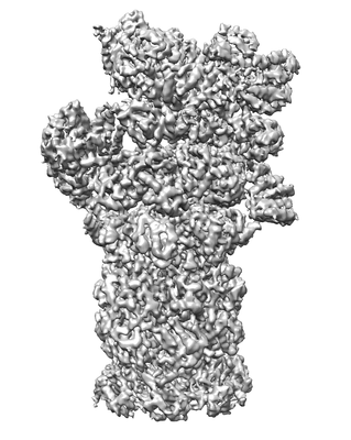 emd_4322.png emd_4322.png | 57.4 KB | ||
| Filedesc metadata | emd-4322.cif.gz | 13.2 KB | ||
| Archive directory |  http://ftp.pdbj.org/pub/emdb/structures/EMD-4322ftp://ftp.pdbj.org/pub/emdb/structures/EMD-4322 http://ftp.pdbj.org/pub/emdb/structures/EMD-4322ftp://ftp.pdbj.org/pub/emdb/structures/EMD-4322 | HTTPS FTP |
-Related structure data
| Related structure data |  6fvwMC  4321C 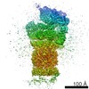 4323C 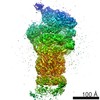 4324C 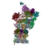 6fvtC 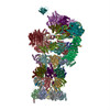 6fvuC 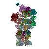 6fvvC  6fvxC 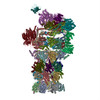 6fvyC M: atomic model generated by this map C: citing same article ( |
|---|---|
| Similar structure data |

-Links
| EMDB pages | EMDB (EBI/PDBe) / EMDataResource |
|---|---|
| Related items in Molecule of the Month |


-Map
| File | Download / File: emd_4322.map.gz / Format: CCP4 / Size: 216 MB / Type: IMAGE STORED AS FLOATING POINT NUMBER (4 BYTES) | ||||||||||||||||||||||||||||||||||||||||||||||||||||||||||||
|---|---|---|---|---|---|---|---|---|---|---|---|---|---|---|---|---|---|---|---|---|---|---|---|---|---|---|---|---|---|---|---|---|---|---|---|---|---|---|---|---|---|---|---|---|---|---|---|---|---|---|---|---|---|---|---|---|---|---|---|---|---|
| Annotation | None | ||||||||||||||||||||||||||||||||||||||||||||||||||||||||||||
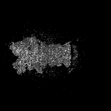
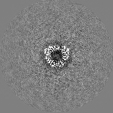
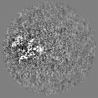
| Projections & slices | Image control
Images are generated by Spider. | ||||||||||||||||||||||||||||||||||||||||||||||||||||||||||||
| Voxel size | X=Y=Z: 1.38 Å | ||||||||||||||||||||||||||||||||||||||||||||||||||||||||||||
| Density |
| ||||||||||||||||||||||||||||||||||||||||||||||||||||||||||||
| Symmetry | Space group: 1 | ||||||||||||||||||||||||||||||||||||||||||||||||||||||||||||
| Details | EMDB XML:
CCP4 map header:
| ||||||||||||||||||||||||||||||||||||||||||||||||||||||||||||
 Z (Sec.)
 Y (Row.)
 X (Col.)


-Supplemental data
- Sample components
+Entire : 26S proteasome
+Supramolecule #1: 26S proteasome
+Macromolecule #1: Proteasome subunit alpha type-1
+Macromolecule #2: Proteasome subunit alpha type-2
+Macromolecule #3: Proteasome subunit alpha type-3
+Macromolecule #4: Proteasome subunit alpha type-4
+Macromolecule #5: Proteasome subunit alpha type-5
+Macromolecule #6: Proteasome subunit alpha type-6
+Macromolecule #7: Probable proteasome subunit alpha type-7
+Macromolecule #8: Proteasome subunit beta type-1
+Macromolecule #9: Proteasome subunit beta type-2
+Macromolecule #10: Proteasome subunit beta type-3
+Macromolecule #11: Proteasome subunit beta type-4
+Macromolecule #12: Proteasome subunit beta type-5
+Macromolecule #13: Proteasome subunit beta type-6
+Macromolecule #14: Proteasome subunit beta type-7
+Macromolecule #15: 26S proteasome regulatory subunit RPN10
+Macromolecule #16: Ubiquitin carboxyl-terminal hydrolase RPN11
+Macromolecule #17: 26S proteasome regulatory subunit RPN12
+Macromolecule #18: 26S proteasome regulatory subunit RPN13
+Macromolecule #19: 26S proteasome complex subunit SEM1
+Macromolecule #20: 26S proteasome regulatory subunit RPN1
+Macromolecule #21: 26S proteasome regulatory subunit RPN2
+Macromolecule #22: 26S proteasome regulatory subunit RPN3
+Macromolecule #23: 26S proteasome regulatory subunit RPN5
+Macromolecule #24: 26S proteasome regulatory subunit RPN6
+Macromolecule #25: 26S proteasome regulatory subunit RPN7
+Macromolecule #26: 26S proteasome regulatory subunit RPN8
+Macromolecule #27: 26S proteasome regulatory subunit RPN9
+Macromolecule #28: 26S proteasome regulatory subunit 7 homolog
+Macromolecule #29: 26S proteasome regulatory subunit 4 homolog
+Macromolecule #30: 26S proteasome regulatory subunit 6B homolog
+Macromolecule #31: 26S proteasome subunit RPT4
+Macromolecule #32: 26S proteasome regulatory subunit 6A
+Macromolecule #33: 26S proteasome regulatory subunit 8 homolog
+Macromolecule #34: ADENOSINE-5'-TRIPHOSPHATE

+Macromolecule #35: MAGNESIUM ION
+Macromolecule #36: ADENOSINE-5'-DIPHOSPHATE

+Macromolecule #37: water

-Experimental details
-Structure determination
| Method | cryo EM |
|---|---|
 Processing Processing | single particle reconstruction |
| Aggregation state | particle |
-Sample preparation
| Buffer | pH: 7.4 |
|---|---|
| Vitrification | Cryogen name: ETHANE-PROPANE |
- Electron microscopy
| Microscope | FEI TITAN KRIOS |
|---|---|
| Image recording | Film or detector model: GATAN K2 SUMMIT (4k x 4k) / Average electron dose: 35.0 e/Å2 |
| Electron beam | Acceleration voltage: 300 kV / Electron source:  FIELD EMISSION GUN FIELD EMISSION GUN |
| Electron optics | Illumination mode: FLOOD BEAM / Imaging mode: BRIGHT FIELD |
| Experimental equipment |  Model: Titan Krios / Image courtesy: FEI Company |
-Image processing
| Startup model | Type of model: PDB ENTRY PDB model - PDB ID: |
|---|---|
| Final reconstruction | Resolution.type: BY AUTHOR / Resolution: 4.5 Å / Resolution method: FSC 0.143 CUT-OFF / Number images used: 351984 |
| Initial angle assignment | Type: PROJECTION MATCHING |
| Final angle assignment | Type: PROJECTION MATCHING |