[English] 日本語
 Yorodumi
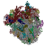
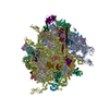
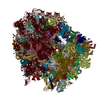
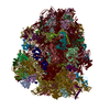
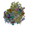
- EMDB-4300: Structure of a prehandover mammalian ribosomal SRP and SRP recept... -
+ Open data
- Basic information
| Entry | Database: EMDB / ID: EMD-4300 | |||||||||
|---|---|---|---|---|---|---|---|---|---|---|
| Title | Structure of a prehandover mammalian ribosomal SRP and SRP receptor targeting complex | |||||||||
 Map data Map data | ||||||||||
 Sample Sample |
| |||||||||
 Keywords Keywords | ER membrane targeting ribosome Signal recognition particle / TRANSLATION | |||||||||
| Function / homology |  Function and homology information Function and homology informationSRP-dependent cotranslational protein targeting to membrane / signal recognition particle receptor complex / SRP-dependent cotranslational protein targeting to membrane, signal sequence recognition / endoplasmic reticulum signal peptide binding / signal recognition particle, endoplasmic reticulum targeting / signal recognition particle binding / granulocyte differentiation / protein targeting to ER / signal-recognition-particle GTPase / protein localization to Golgi apparatus ...SRP-dependent cotranslational protein targeting to membrane / signal recognition particle receptor complex / SRP-dependent cotranslational protein targeting to membrane, signal sequence recognition / endoplasmic reticulum signal peptide binding / signal recognition particle, endoplasmic reticulum targeting / signal recognition particle binding / granulocyte differentiation / protein targeting to ER / signal-recognition-particle GTPase / protein localization to Golgi apparatus / negative regulation of translational elongation / SRP-dependent cotranslational protein targeting to membrane, translocation / 7S RNA binding / Golgi to plasma membrane protein transport / SRP-dependent cotranslational protein targeting to membrane / exocrine pancreas development / TPR domain binding / ribonucleoprotein complex binding / cytoplasmic microtubule / neutrophil chemotaxis / intracellular protein transport / GDP binding / ribosome binding / nuclear speck / GTPase activity / endoplasmic reticulum membrane / GTP binding / nucleolus / endoplasmic reticulum / Golgi apparatus / ATP hydrolysis activity / nucleoplasm / nucleus / cytosol Similarity search - Function | |||||||||
| Biological species |    | |||||||||
| Method | single particle reconstruction / cryo EM / Resolution: 3.7 Å | |||||||||
 Authors Authors | Kobayashi K / Jomaa A | |||||||||
| Funding support |  Switzerland, 2 items Switzerland, 2 items
| |||||||||
 Citation Citation |  Journal: Science / Year: 2018 Journal: Science / Year: 2018Title: Structure of a prehandover mammalian ribosomal SRP·SRP receptor targeting complex. Authors: Kan Kobayashi / Ahmad Jomaa / Jae Ho Lee / Sowmya Chandrasekar / Daniel Boehringer / Shu-Ou Shan / Nenad Ban /   Abstract: Signal recognition particle (SRP) targets proteins to the endoplasmic reticulum (ER). SRP recognizes the ribosome synthesizing a signal sequence and delivers it to the SRP receptor (SR) on the ER ...Signal recognition particle (SRP) targets proteins to the endoplasmic reticulum (ER). SRP recognizes the ribosome synthesizing a signal sequence and delivers it to the SRP receptor (SR) on the ER membrane followed by the transfer of the signal sequence to the translocon. Here, we present the cryo-electron microscopy structure of the mammalian translating ribosome in complex with SRP and SR in a conformation preceding signal sequence handover. The structure visualizes all eukaryotic-specific SRP and SR proteins and reveals their roles in stabilizing this conformation by forming a large protein assembly at the distal site of SRP RNA. We provide biochemical evidence that the guanosine triphosphate hydrolysis of SRP·SR is delayed at this stage, possibly to provide a time window for signal sequence handover to the translocon. | |||||||||
| History |
|
- Structure visualization
| Movie |
 Movie viewer Movie viewer |
|---|---|
| Structure viewer | EM map:  SurfView SurfView Molmil Molmil Jmol/JSmol Jmol/JSmol |
- Downloads & links
-EMDB archive
| Map data |  emd_4300.map.gz emd_4300.map.gz | 116.3 MB |  EMDB map data format EMDB map data format | |
|---|---|---|---|---|
| Header (meta data) |  emd-4300-v30.xml emd-4300-v30.xml emd-4300.xml emd-4300.xml | 89 KB 89 KB | Display Display |  EMDB header EMDB header |
| Images |  emd_4300.png emd_4300.png | 265.7 KB | ||
| Filedesc metadata |  emd-4300.cif.gz emd-4300.cif.gz | 17.2 KB | ||
| Others |  emd_4300_half_map_1.map.gz emd_4300_half_map_1.map.gz emd_4300_half_map_2.map.gz emd_4300_half_map_2.map.gz | 98 MB 98.2 MB | ||
| Archive directory |  http://ftp.pdbj.org/pub/emdb/structures/EMD-4300 http://ftp.pdbj.org/pub/emdb/structures/EMD-4300 ftp://ftp.pdbj.org/pub/emdb/structures/EMD-4300 ftp://ftp.pdbj.org/pub/emdb/structures/EMD-4300 | HTTPS FTP |
-Validation report
| Summary document |  emd_4300_validation.pdf.gz emd_4300_validation.pdf.gz | 1 MB | Display |  EMDB validaton report EMDB validaton report |
|---|---|---|---|---|
| Full document |  emd_4300_full_validation.pdf.gz emd_4300_full_validation.pdf.gz | 1 MB | Display | |
| Data in XML |  emd_4300_validation.xml.gz emd_4300_validation.xml.gz | 14 KB | Display | |
| Data in CIF |  emd_4300_validation.cif.gz emd_4300_validation.cif.gz | 16.5 KB | Display | |
| Arichive directory |  https://ftp.pdbj.org/pub/emdb/validation_reports/EMD-4300 https://ftp.pdbj.org/pub/emdb/validation_reports/EMD-4300 ftp://ftp.pdbj.org/pub/emdb/validation_reports/EMD-4300 ftp://ftp.pdbj.org/pub/emdb/validation_reports/EMD-4300 | HTTPS FTP |
-Related structure data
| Related structure data |  6frkMC M: atomic model generated by this map C: citing same article ( |
|---|---|
| Similar structure data |
- Links
Links
| EMDB pages |  EMDB (EBI/PDBe) / EMDB (EBI/PDBe) /  EMDataResource EMDataResource |
|---|---|
| Related items in Molecule of the Month |
- Map
Map
| File |  Download / File: emd_4300.map.gz / Format: CCP4 / Size: 125 MB / Type: IMAGE STORED AS FLOATING POINT NUMBER (4 BYTES) Download / File: emd_4300.map.gz / Format: CCP4 / Size: 125 MB / Type: IMAGE STORED AS FLOATING POINT NUMBER (4 BYTES) | ||||||||||||||||||||||||||||||||||||||||||||||||||||||||||||
|---|---|---|---|---|---|---|---|---|---|---|---|---|---|---|---|---|---|---|---|---|---|---|---|---|---|---|---|---|---|---|---|---|---|---|---|---|---|---|---|---|---|---|---|---|---|---|---|---|---|---|---|---|---|---|---|---|---|---|---|---|---|
| Voxel size | X=Y=Z: 1.39 Å | ||||||||||||||||||||||||||||||||||||||||||||||||||||||||||||
| Density |
| ||||||||||||||||||||||||||||||||||||||||||||||||||||||||||||
| Symmetry | Space group: 1 | ||||||||||||||||||||||||||||||||||||||||||||||||||||||||||||
| Details | EMDB XML:
CCP4 map header:
| ||||||||||||||||||||||||||||||||||||||||||||||||||||||||||||
-Supplemental data
- Sample components
+Entire : Translating ribosome bound to SRP and SRP receptor
+Supramolecule #1: Translating ribosome bound to SRP and SRP receptor
+Supramolecule #2: Ribosome
+Supramolecule #3: SRP
+Supramolecule #4: SRP receptor
+Supramolecule #5: Signal sequence
+Macromolecule #1: Canis lupus familiaris RNA, 7SL, cytoplasmic 1 (RN7SL1), SRP RNA
+Macromolecule #2: tRNA
+Macromolecule #5: 28S ribosomal RNA
+Macromolecule #7: 5S ribosomal RNA
+Macromolecule #8: 5.8S ribosomal RNA
+Macromolecule #3: Ribosomal protein eL28
+Macromolecule #4: Ribosomal protein L12
+Macromolecule #6: 60S acidic ribosomal protein P0
+Macromolecule #9: Ribosomal protein L8
+Macromolecule #10: Ribosomal protein uL3
+Macromolecule #11: Ribosomal protein uL4
+Macromolecule #12: Ribosomal protein uL18
+Macromolecule #13: Ribosomal protein eL6
+Macromolecule #14: Ribosomal protein uL30
+Macromolecule #15: Ribosomal protein eL8
+Macromolecule #16: Ribosomal protein uL6
+Macromolecule #17: Ribosomal protein uL16
+Macromolecule #18: Ribosomal protein uL5
+Macromolecule #19: Ribosomal protein eL13
+Macromolecule #20: Ribosomal protein eL14
+Macromolecule #21: Ribosomal protein L15
+Macromolecule #22: Ribosomal protein uL13
+Macromolecule #23: Ribosomal protein uL22
+Macromolecule #24: Ribosomal protein eL18
+Macromolecule #25: Ribosomal protein eL19
+Macromolecule #26: Ribosomal protein eL20
+Macromolecule #27: Ribosomal protein eL21
+Macromolecule #28: Ribosomal protein eL22
+Macromolecule #29: Ribosomal protein L23
+Macromolecule #30: Ribosomal protein eL24
+Macromolecule #31: Ribosomal protein uL23
+Macromolecule #32: Ribosomal protein uL24
+Macromolecule #33: 60S ribosomal protein L27
+Macromolecule #34: Ribosomal protein uL15
+Macromolecule #35: 60S ribosomal protein L29
+Macromolecule #36: Ribosomal protein eL30
+Macromolecule #37: Ribosomal protein eL31
+Macromolecule #38: Ribosomal protein eL32
+Macromolecule #39: Ribosomal protein eL33
+Macromolecule #40: Ribosomal protein eL34
+Macromolecule #41: Ribosomal protein uL29
+Macromolecule #42: Ribosomal protein eL36
+Macromolecule #43: Ribosomal protein L37
+Macromolecule #44: Ribosomal protein eL38
+Macromolecule #45: Ribosomal protein eL39
+Macromolecule #46: Ribosomal protein eL40
+Macromolecule #47: Ribosomal protein eL41
+Macromolecule #48: Ribosomal protein eL42
+Macromolecule #49: Ribosomal protein eL43
+Macromolecule #50: Signal recognition particle subunit SRP19
+Macromolecule #51: Signal recognition particle subunit SRP72,Signal recognition part...
+Macromolecule #52: Signal sequence
+Macromolecule #53: Signal recognition particle subunit SRP68
+Macromolecule #54: SRP receptor beta subunit
+Macromolecule #55: Signal recognition particle 9 kDa protein
+Macromolecule #56: Signal recognition particle 54 kDa protein
+Macromolecule #57: SRP receptor alpha subunit
+Macromolecule #58: Signal recognition particle 14 kDa protein
+Macromolecule #59: MAGNESIUM ION
+Macromolecule #60: ZINC ION
+Macromolecule #61: GUANOSINE-5'-TRIPHOSPHATE
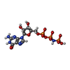
+Macromolecule #62: PHOSPHOAMINOPHOSPHONIC ACID-GUANYLATE ESTER
-Experimental details
-Structure determination
| Method | cryo EM |
|---|---|
 Processing Processing | single particle reconstruction |
| Aggregation state | particle |
- Sample preparation
| Buffer | pH: 7.6 |
|---|---|
| Grid | Model: Quantifoil R2/2 / Material: COPPER / Support film - Material: CARBON |
| Vitrification | Cryogen name: ETHANE-PROPANE / Chamber humidity: 100 % / Chamber temperature: 277 K / Instrument: FEI VITROBOT MARK IV |
- Electron microscopy
| Microscope | FEI TITAN KRIOS |
|---|---|
| Image recording | Film or detector model: FEI FALCON II (4k x 4k) / Detector mode: INTEGRATING / Average electron dose: 40.0 e/Å2 |
| Electron beam | Acceleration voltage: 300 kV / Electron source:  FIELD EMISSION GUN FIELD EMISSION GUN |
| Electron optics | Illumination mode: FLOOD BEAM / Imaging mode: BRIGHT FIELD / Cs: 2.7 mm / Nominal magnification: 59000 |
| Sample stage | Specimen holder model: FEI TITAN KRIOS AUTOGRID HOLDER |
| Experimental equipment |  Model: Titan Krios / Image courtesy: FEI Company |
+ Image processing
-Atomic model buiding 1
| Refinement | Space: RECIPROCAL / Protocol: RIGID BODY FIT |
|---|---|
| Output model |  PDB-6frk: |
 Movie
Movie Controller


