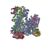
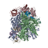
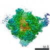
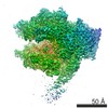
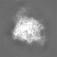
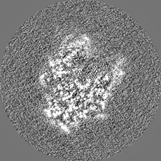
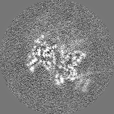
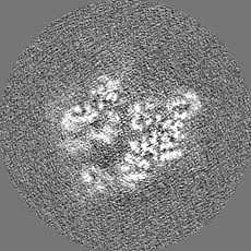
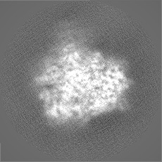
- EMDB-4147: Yeast RNA polymerase I elongation complex at 3.8A -
+
Open data
ID or keywords:
Loading...
-
Basic information
Entry
Database: EMDB / ID: EMD-4147
Title
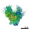
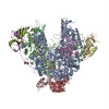
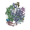
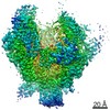
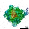
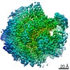
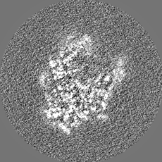
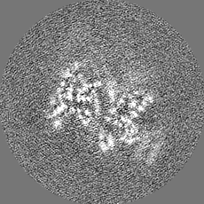
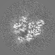
Yeast RNA polymerase I elongation complex at 3.8A
Map data
Sharpened map (B=-149A^2)
Sample
Complex: Yeast RNA polymerase I elongation complex
Protein or peptide: x 14 types
DNA: x 2 types
RNA: x 1 types
Ligand: x 3 types
Keywords
RNA polymerase I / transcription
Function / homology
Function and homology information
nuclear DNA-directed RNA polymerase complex / RNA polymerase I preinitiation complex assembly / RNA Polymerase I Transcription Initiation / regulation of cell size / Processing of Capped Intron-Containing Pre-mRNA / RNA Polymerase III Transcription Initiation From Type 2 Promoter / RNA Pol II CTD phosphorylation and interaction with CE / Formation of the Early Elongation Complex / mRNA Capping / RNA polymerase II transcribes snRNA genes ...nuclear DNA-directed RNA polymerase complex / RNA polymerase I preinitiation complex assembly / RNA Polymerase I Transcription Initiation / regulation of cell size / Processing of Capped Intron-Containing Pre-mRNA / RNA Polymerase III Transcription Initiation From Type 2 Promoter / RNA Pol II CTD phosphorylation and interaction with CE / Formation of the Early Elongation Complex / mRNA Capping / RNA polymerase II transcribes snRNA genes / TP53 Regulates Transcription of DNA Repair Genes / RNA Polymerase II Promoter Escape / RNA Polymerase II Transcription Pre-Initiation And Promoter Opening / RNA Polymerase II Transcription Initiation / RNA Polymerase II Transcription Initiation And Promoter Clearance / RNA Polymerase II Pre-transcription Events / Formation of TC-NER Pre-Incision Complex / RNA-templated transcription / RNA Polymerase I Promoter Escape / termination of RNA polymerase III transcription / termination of RNA polymerase I transcription / transcription initiation at RNA polymerase III promoter / Gap-filling DNA repair synthesis and ligation in TC-NER / nucleolar large rRNA transcription by RNA polymerase I / transcription initiation at RNA polymerase I promoter / Estrogen-dependent gene expression / transcription by RNA polymerase III / Dual incision in TC-NER / RNA polymerase I complex / RNA polymerase III complex / transcription elongation by RNA polymerase I / RNA polymerase II, core complex / tRNA transcription by RNA polymerase III / transcription by RNA polymerase I / promoter-specific chromatin binding / transcription initiation at RNA polymerase II promoter / transcription elongation by RNA polymerase II / ribonucleoside binding / DNA-directed RNA polymerase / DNA-directed RNA polymerase activity / peroxisome / ribosome biogenesis / transcription by RNA polymerase II / nucleic acid binding / RNA polymerase II-specific DNA-binding transcription factor binding / protein dimerization activity / nucleolus / negative regulation of transcription by RNA polymerase II / DNA binding / zinc ion binding / nucleoplasm / metal ion binding / nucleus / cytoplasm Similarity search - Function
DNA-directed RNA polymerases I and III subunit RPAC1 / DNA-directed RNA polymerase I subunit RPA190 / DNA-directed RNA polymerases I, II, and III subunit RPABC1 / DNA-directed RNA polymerases I, II, and III subunit RPABC2 / DNA-directed RNA polymerases I, II, and III subunit RPABC3 / DNA-directed RNA polymerase I subunit RPA135 / DNA-directed RNA polymerases I, II, and III subunit RPABC5 / DNA-directed RNA polymerases I and III subunit RPAC2 / DNA-directed RNA polymerase I subunit RPA12 / DNA-directed RNA polymerases I, II, and III subunit RPABC4 ...DNA-directed RNA polymerases I and III subunit RPAC1 / DNA-directed RNA polymerase I subunit RPA190 / DNA-directed RNA polymerases I, II, and III subunit RPABC1 / DNA-directed RNA polymerases I, II, and III subunit RPABC2 / DNA-directed RNA polymerases I, II, and III subunit RPABC3 / DNA-directed RNA polymerase I subunit RPA135 / DNA-directed RNA polymerases I, II, and III subunit RPABC5 / DNA-directed RNA polymerases I and III subunit RPAC2 / DNA-directed RNA polymerase I subunit RPA12 / DNA-directed RNA polymerases I, II, and III subunit RPABC4 / DNA-directed RNA polymerase I subunit RPA43 / DNA-directed RNA polymerase I subunit RPA34 / DNA-directed RNA polymerase I subunit RPA14 / DNA-directed RNA polymerase I subunit RPA49 Similarity search - Component
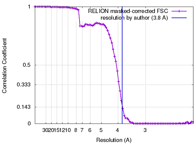
Journal: Nature / Year: 2016 Title: Structure of RNA polymerase I transcribing ribosomal DNA genes. Authors: Simon Neyer / Michael Kunz / Christian Geiss / Merle Hantsche / Victor-Valentin Hodirnau / Anja Seybert / Christoph Engel / Margot P Scheffer / Patrick Cramer / Achilleas S Frangakis / Abstract: RNA polymerase I (Pol I) is a highly processive enzyme that transcribes ribosomal DNA (rDNA) and regulates growth of eukaryotic cells. Crystal structures of free Pol I from the yeast Saccharomyces ...RNA polymerase I (Pol I) is a highly processive enzyme that transcribes ribosomal DNA (rDNA) and regulates growth of eukaryotic cells. Crystal structures of free Pol I from the yeast Saccharomyces cerevisiae have revealed dimers of the enzyme stabilized by a 'connector' element and an expanded cleft containing the active centre in an inactive conformation. The central bridge helix was unfolded and a Pol-I-specific 'expander' element occupied the DNA-template-binding site. The structure of Pol I in its active transcribing conformation has yet to be determined, whereas structures of Pol II and Pol III have been solved with bound DNA template and RNA transcript. Here we report structures of active transcribing Pol I from yeast solved by two different cryo-electron microscopy approaches. A single-particle structure at 3.8 Å resolution reveals a contracted active centre cleft with bound DNA and RNA, and a narrowed pore beneath the active site that no longer holds the RNA-cleavage-stimulating domain of subunit A12.2. A structure at 29 Å resolution that was determined from cryo-electron tomograms of Pol I enzymes transcribing cellular rDNA confirms contraction of the cleft and reveals that incoming and exiting rDNA enclose an angle of around 150°. The structures suggest a model for the regulation of transcription elongation in which contracted and expanded polymerase conformations are associated with active and inactive states, respectively.
History
Deposition
Oct 14, 2016
-
Header (metadata) release
Nov 23, 2016
-
Map release
Nov 23, 2016
-
Update
Dec 17, 2025
-
Current status
Dec 17, 2025
Processing site: PDBe / Status: Released
-
Structure visualization
Movie
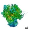
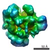
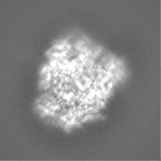
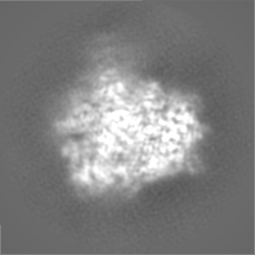
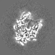
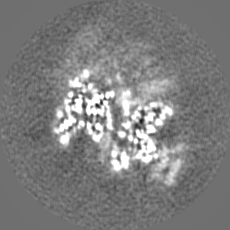
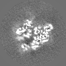
Surface view with section colored by density value
Cryogen name: ETHANE / Chamber humidity: 100 % / Chamber temperature: 277 K / Instrument: FEI VITROBOT MARK IV / Details: blot force 13, blotting time 8.5s.
-
Electron microscopy
Microscope
FEI TITAN KRIOS
Image recording
Film or detector model: GATAN K2 QUANTUM (4k x 4k) / Average electron dose: 56.0 e/Å2
Electron beam
Acceleration voltage: 300 kV / Electron source: FIELD EMISSION GUN
Electron optics
Illumination mode: FLOOD BEAM / Imaging mode: BRIGHT FIELD
In the structure databanks used in Yorodumi, some data are registered as the other names, "COVID-19 virus" and "2019-nCoV". Here are the details of the virus and the list of structure data.
Jan 31, 2019. EMDB accession codes are about to change! (news from PDBe EMDB page)
EMDB accession codes are about to change! (news from PDBe EMDB page)
The allocation of 4 digits for EMDB accession codes will soon come to an end. Whilst these codes will remain in use, new EMDB accession codes will include an additional digit and will expand incrementally as the available range of codes is exhausted. The current 4-digit format prefixed with “EMD-” (i.e. EMD-XXXX) will advance to a 5-digit format (i.e. EMD-XXXXX), and so on. It is currently estimated that the 4-digit codes will be depleted around Spring 2019, at which point the 5-digit format will come into force.
The EM Navigator/Yorodumi systems omit the EMD- prefix.
Related info.:Q: What is EMD? / ID/Accession-code notation in Yorodumi/EM Navigator
Yorodumi is a browser for structure data from EMDB, PDB, SASBDB, etc.
This page is also the successor to EM Navigator detail page, and also detail information page/front-end page for Omokage search.
The word "yorodu" (or yorozu) is an old Japanese word meaning "ten thousand". "mi" (miru) is to see.
Related info.:EMDB / PDB / SASBDB / Comparison of 3 databanks / Yorodumi Search / Aug 31, 2016. New EM Navigator & Yorodumi / Yorodumi Papers / Jmol/JSmol / Function and homology information / Changes in new EM Navigator and Yorodumi
 Movie
Movie Controller


 Open data
 Basic information
 Map data
 Sample
 Keywords
 Function and homology information
 Authors
 Citation
 Structure visualization
 Downloads & links
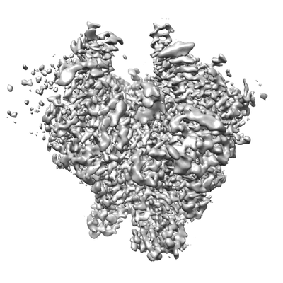 emd_4147.png
 http://ftp.pdbj.org/pub/emdb/structures/EMD-4147
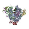


 Z (Sec.)
 Y (Row.)
 X (Col.)


 Sample components
 Processing
 Electron microscopy
 FIELD EMISSION GUN