 Movie
Movie Controller


+ Open data

- Basic information
| Entry |  | |||||||||
|---|---|---|---|---|---|---|---|---|---|---|
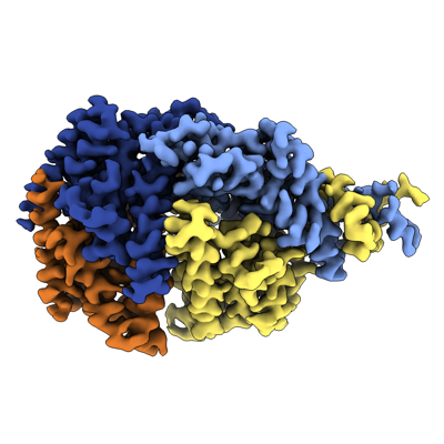
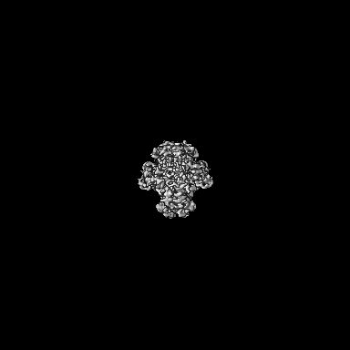
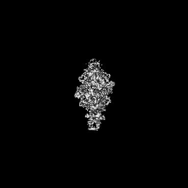
| Title | CryoEM structure of Shedu from Bacillus cereus | |||||||||
 Map data Map data | CryoEM structure of Shedu from Bacillus cereus | |||||||||
 Sample Sample |
| |||||||||
 Keywords Keywords | Shedu / DUF4263 / Bacterial defense systems / Nuclease / Anti-plasmid defense system / PD-(D/E)XK nuclease / Whirly domain / Two-component signaling / DNA BINDING PROTEIN | |||||||||
| Function / homology | Protein of unknown function DUF4263 / : / Shedu protein SduA, C-terminal / Shedu protein SduA, N-terminal / nuclease activity / defense response to virus / Shedu protein SduA Function and homology information Function and homology information | |||||||||
| Biological species |  | |||||||||
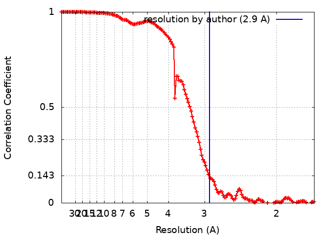
| Method | single particle reconstruction / cryo EM / Resolution: 2.9 Å | |||||||||
 Authors Authors | Gu Y / Corbett K | |||||||||
| Funding support |  United States, 1 items United States, 1 items
| |||||||||
 Citation Citation | Journal: to be published Title: Shedu anti-phage nucleases share a common enzymatic core regulated by diverse sensor domains Authors: Gu Y / Corbett K | |||||||||
| History |
|
- Structure visualization
| Supplemental images |
|---|
- Downloads & links
-EMDB archive
| Map data | emd_41281.map.gz | 108.3 MB | EMDB map data format | |
|---|---|---|---|---|
| Header (meta data) | emd-41281-v30.xmlemd-41281.xml | 15.2 KB 15.2 KB | Display Display | EMDB header |
| FSC (resolution estimation) | emd_41281_fsc.xml | 13.3 KB | Display | FSC data file |
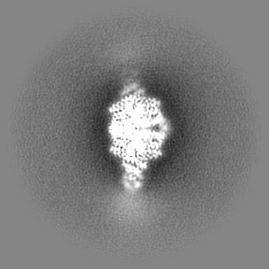
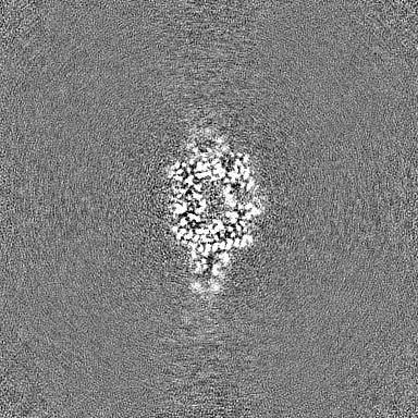
| Images |  emd_41281.png emd_41281.png | 113.2 KB | ||
| Filedesc metadata | emd-41281.cif.gz | 5.8 KB | ||
| Others | emd_41281_half_map_1.map.gzemd_41281_half_map_2.map.gz | 200.7 MB 200.7 MB | ||
| Archive directory |  http://ftp.pdbj.org/pub/emdb/structures/EMD-41281ftp://ftp.pdbj.org/pub/emdb/structures/EMD-41281 http://ftp.pdbj.org/pub/emdb/structures/EMD-41281ftp://ftp.pdbj.org/pub/emdb/structures/EMD-41281 | HTTPS FTP |
-Validation report
| Summary document | emd_41281_validation.pdf.gz | 897.8 KB | Display | EMDB validaton report |
|---|---|---|---|---|
| Full document | emd_41281_full_validation.pdf.gz | 897.3 KB | Display | |
| Data in XML | emd_41281_validation.xml.gz | 21.8 KB | Display | |
| Data in CIF | emd_41281_validation.cif.gz | 28 KB | Display | |
| Arichive directory | https://ftp.pdbj.org/pub/emdb/validation_reports/EMD-41281ftp://ftp.pdbj.org/pub/emdb/validation_reports/EMD-41281 | HTTPS FTP |
-Related structure data
| Related structure data | 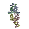 8ti8MC 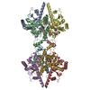 8ti9C 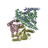 8tiaC M: atomic model generated by this map C: citing same article ( |
|---|---|
| Similar structure data |

-Links
| EMDB pages | EMDB (EBI/PDBe) / EMDataResource |
|---|
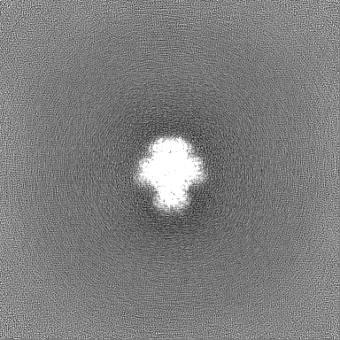
-Map
| File | Download / File: emd_41281.map.gz / Format: CCP4 / Size: 216 MB / Type: IMAGE STORED AS FLOATING POINT NUMBER (4 BYTES) | ||||||||||||||||||||||||||||||||||||
|---|---|---|---|---|---|---|---|---|---|---|---|---|---|---|---|---|---|---|---|---|---|---|---|---|---|---|---|---|---|---|---|---|---|---|---|---|---|
| Annotation | CryoEM structure of Shedu from Bacillus cereus | ||||||||||||||||||||||||||||||||||||
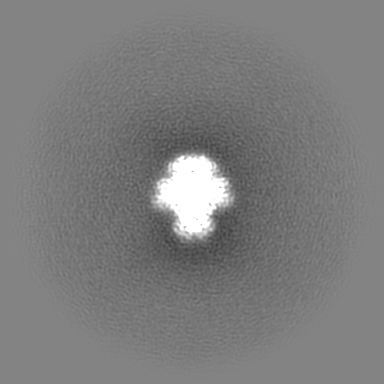
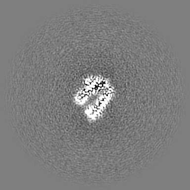
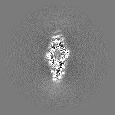
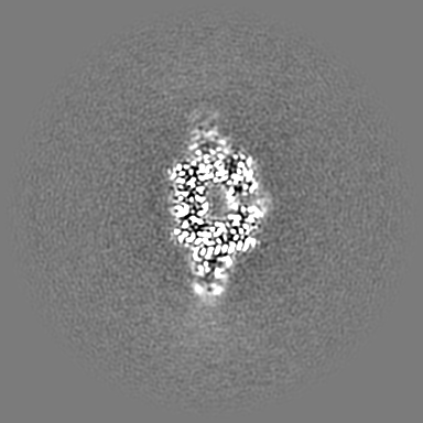
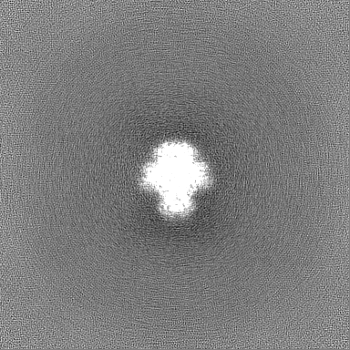
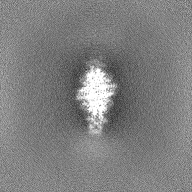
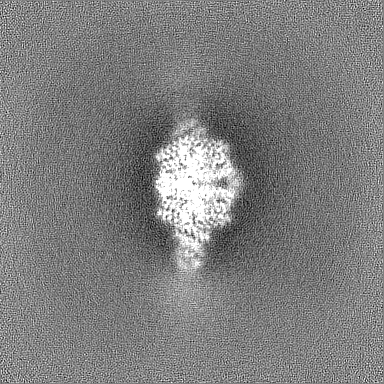
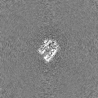
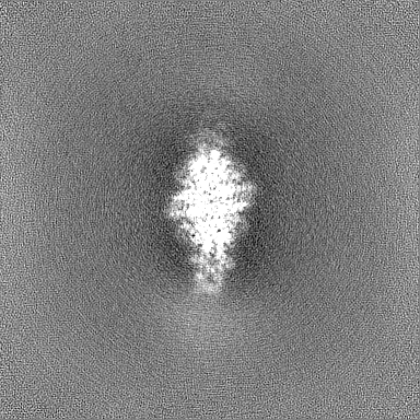
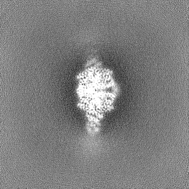
| Projections & slices | Image control
Images are generated by Spider. | ||||||||||||||||||||||||||||||||||||
| Voxel size | X=Y=Z: 0.84 Å | ||||||||||||||||||||||||||||||||||||
| Density |
| ||||||||||||||||||||||||||||||||||||
| Symmetry | Space group: 1 | ||||||||||||||||||||||||||||||||||||
| Details | EMDB XML:
|
 Z (Sec.)
 Y (Row.)
 X (Col.)


-Supplemental data
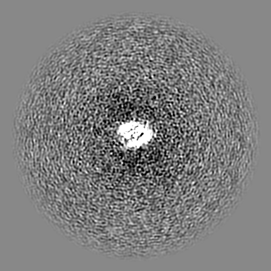
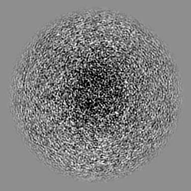
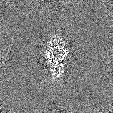
-Half map: Half Map 1
| File | emd_41281_half_map_1.map | ||||||||||||
|---|---|---|---|---|---|---|---|---|---|---|---|---|---|
| Annotation | Half Map 1 | ||||||||||||
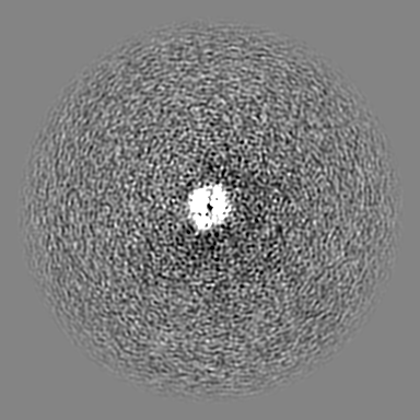
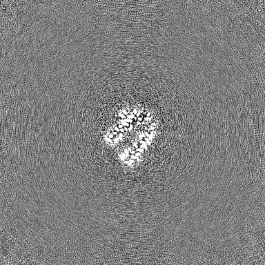
| Projections & Slices |
| ||||||||||||
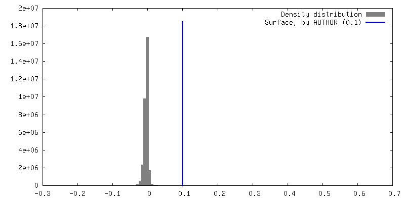
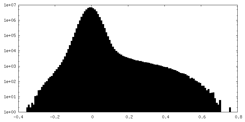
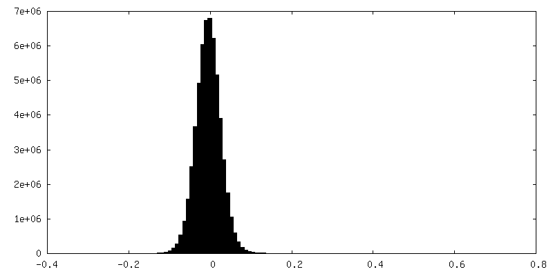
| Density Histograms |
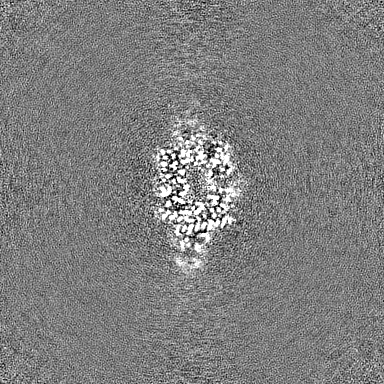

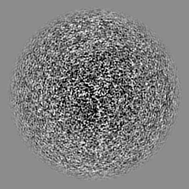
-Half map: Half Map 2
| File | emd_41281_half_map_2.map | ||||||||||||
|---|---|---|---|---|---|---|---|---|---|---|---|---|---|
| Annotation | Half Map 2 | ||||||||||||
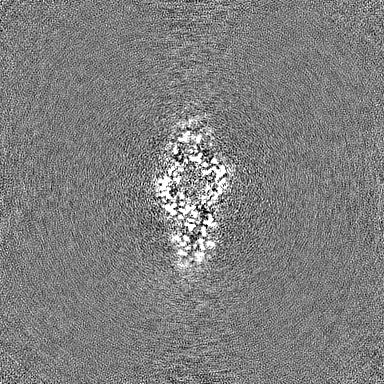
| Projections & Slices |
| ||||||||||||
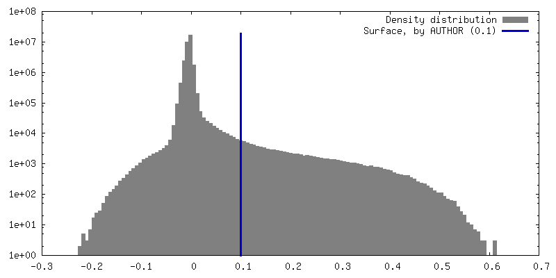
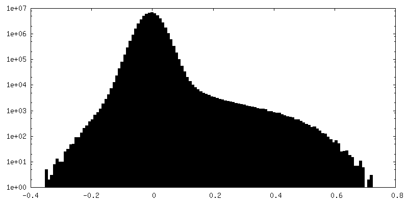
| Density Histograms |
- Sample components
-Entire : Bacillus cereus Shedu Tetramer
| Entire | Name: Bacillus cereus Shedu Tetramer |
|---|---|
| Components |
|
-Supramolecule #1: Bacillus cereus Shedu Tetramer
| Supramolecule | Name: Bacillus cereus Shedu Tetramer / type: complex / ID: 1 / Parent: 0 / Macromolecule list: all |
|---|---|
| Source (natural) | Organism: |
| Molecular weight | Theoretical: 175 KDa |
-Macromolecule #1: Shedu protein SduA
| Macromolecule | Name: Shedu protein SduA / type: protein_or_peptide / ID: 1 / Number of copies: 4 / Enantiomer: LEVO |
|---|---|
| Source (natural) | Organism: |
| Molecular weight | Theoretical: 44.016969 KDa |
| Recombinant expression | Organism: |
| Sequence | String: SNASDKINVW TTSRDSAVCG DIELKKTSTT RLIFRPEIVN NNKNPKASVR GCFIFQKKGR NALWDDYKEL DMNKLKAEEW IKLEINSDA MLTLTKEIQK HYAVHEKYGV RYGAFHLFKD NPDIEKLIEM FESNTDLLTQ LMEDDKSEAL EKTLEWIVTN D NPDKIIDR ...String: SNASDKINVW TTSRDSAVCG DIELKKTSTT RLIFRPEIVN NNKNPKASVR GCFIFQKKGR NALWDDYKEL DMNKLKAEEW IKLEINSDA MLTLTKEIQK HYAVHEKYGV RYGAFHLFKD NPDIEKLIEM FESNTDLLTQ LMEDDKSEAL EKTLEWIVTN D NPDKIIDR LKNLKEQDLD QLNTLIGIAN LKKVLSVWES NKLTNTSEKF WQSVLKENTW ILSQIFSNPT VLINDEAYVG GK TVKNDSG KLVDFLYANP FSKDAVLIEI KTPSTPLITP TEYRTGVYSA HKDLTGAVTQ VLTYKTTLQR EYQNIDYNNY RQG IKTDFD IITPCCVVIA GMFDTLTDTA HRHSFELYRK ELKNVTVITF DELFERVKGL IKLLEG UniProtKB: Shedu protein SduA |
-Experimental details
-Structure determination
| Method | cryo EM |
|---|---|
 Processing Processing | single particle reconstruction |
| Aggregation state | particle |
-Sample preparation
| Concentration | 0.79 mg/mL | |||||||||||||||
|---|---|---|---|---|---|---|---|---|---|---|---|---|---|---|---|---|
| Buffer | pH: 8.5 Component:
Details: Prepared using deionized water and filtered strelized. | |||||||||||||||
| Vitrification | Cryogen name: ETHANE / Chamber humidity: 100 % / Chamber temperature: 277 K / Instrument: FEI VITROBOT MARK IV | |||||||||||||||
| Details | Freshly collected from size-exclusion column |
- Electron microscopy
| Microscope | FEI TITAN KRIOS |
|---|---|
| Image recording | Film or detector model: GATAN K2 QUANTUM (4k x 4k) / Detector mode: COUNTING / Average electron dose: 55.0 e/Å2 |
| Electron beam | Acceleration voltage: 300 kV / Electron source:  FIELD EMISSION GUN FIELD EMISSION GUN |
| Electron optics | Illumination mode: FLOOD BEAM / Imaging mode: BRIGHT FIELD / Nominal defocus max: 2.0 µm / Nominal defocus min: 0.8 µm |
| Experimental equipment |  Model: Titan Krios / Image courtesy: FEI Company |
+Image processing
-Atomic model buiding 1
| Refinement | Space: REAL / Protocol: AB INITIO MODEL |
|---|---|
| Output model | PDB-8ti8: |
