 Movie
Movie Controller

[English] 日本語
 Yorodumi
- PDB-3jch: Cryo-EM structure of the magnesium channel CorA in the magnesium-... -
+ Open data

- Basic information
| Entry | Database: PDB / ID: 3jch | ||||||
|---|---|---|---|---|---|---|---|
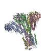
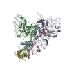
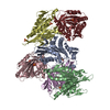
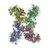
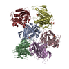
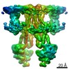
| Title | Cryo-EM structure of the magnesium channel CorA in the magnesium-free, asymmetric open state II | ||||||
 Components Components | Magnesium transport protein CorA | ||||||
 Keywords Keywords | TRANSPORT PROTEIN / membrane protein / ion channel / magnesium channel / pentameric complex / symmetry vs. asymmetry / conformational change / gating mechanism / direct electron detector / K2 | ||||||
| Function / homology |  Function and homology information Function and homology informationcobalt ion transport / cobalt ion transmembrane transporter activity / magnesium ion transmembrane transport / magnesium ion transmembrane transporter activity / cobalt ion binding / protein homooligomerization / magnesium ion binding / identical protein binding / plasma membrane Similarity search - Function | ||||||
| Biological species |   Thermotoga maritima (bacteria) Thermotoga maritima (bacteria) | ||||||
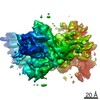
| Method | ELECTRON MICROSCOPY / single particle reconstruction / cryo EM / Resolution: 7.06 Å | ||||||
 Authors Authors | Matthies, D. / Perozo, E. / Subramaniam, S. | ||||||
 Citation Citation | Journal: Cell / Year: 2016 Title: Cryo-EM Structures of the Magnesium Channel CorA Reveal Symmetry Break upon Gating. Authors: Doreen Matthies / Olivier Dalmas / Mario J Borgnia / Pawel K Dominik / Alan Merk / Prashant Rao / Bharat G Reddy / Shahidul Islam / Alberto Bartesaghi / Eduardo Perozo / Sriram Subramaniam /  Abstract: CorA, the major Mg(2+) uptake system in prokaryotes, is gated by intracellular Mg(2+) (KD ∼ 1-2 mM). X-ray crystallographic studies of CorA show similar conformations under Mg(2+)-bound and Mg(2+)- ...CorA, the major Mg(2+) uptake system in prokaryotes, is gated by intracellular Mg(2+) (KD ∼ 1-2 mM). X-ray crystallographic studies of CorA show similar conformations under Mg(2+)-bound and Mg(2+)-free conditions, but EPR spectroscopic studies reveal large Mg(2+)-driven quaternary conformational changes. Here, we determined cryo-EM structures of CorA in the Mg(2+)-bound closed conformation and in two open Mg(2+)-free states at resolutions of 3.8, 7.1, and 7.1 Å, respectively. In the absence of bound Mg(2+), four of the five subunits are displaced to variable extents (∼ 10-25 Å) by hinge-like motions as large as ∼ 35° at the stalk helix. The transition between a single 5-fold symmetric closed state and an ensemble of low Mg(2+), open, asymmetric conformational states is, thus, the key structural signature of CorA gating. This mechanism is likely to apply to other structurally similar divalent ion channels. | ||||||
| History |
|
- Structure visualization
| Movie |
Movie viewer |
|---|---|
| Structure viewer | Molecule: MolmilJmol/JSmol |
- Downloads & links
-Download
| PDBx/mmCIF format | 3jch.cif.gz | 231.1 KB | Display | PDBx/mmCIF format |
|---|---|---|---|---|
| PDB format | pdb3jch.ent.gz | 139.4 KB | Display | PDB format |
| PDBx/mmJSON format | 3jch.json.gz | Tree view | PDBx/mmJSON format | |
| Others |  Other downloads Other downloads |
-Validation report
| Arichive directory | https://data.pdbj.org/pub/pdb/validation_reports/jc/3jchftp://data.pdbj.org/pub/pdb/validation_reports/jc/3jch | HTTPS FTP |
|---|
-Related structure data
| Related structure data | 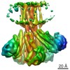 6553MC 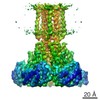 6551C 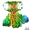 6552C 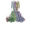 3jcfC 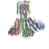 3jcgC M: map data used to model this data C: citing same article ( |
|---|---|
| Similar structure data |
-Links
 PDBj
PDBj- Assembly
| Deposited unit | 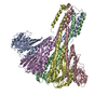
|
|---|---|
| 1 |
|
-Components
| #1: Protein | Mass: 41498.082 Da / Num. of mol.: 5 Source method: isolated from a genetically manipulated source Source: (gene. exp.) Thermotoga maritima (bacteria) / Gene: corA, TM_0561 / Plasmid: CorA-pET15b / Production host: |
|---|
-Experimental details
-Experiment
| Experiment | Method: ELECTRON MICROSCOPY |
|---|---|
| EM experiment | Aggregation state: PARTICLE / 3D reconstruction method: single particle reconstruction |
- Sample preparation
| Component | Name: CorA from Thermotoga maritima in the absence of magnesium, state II Type: COMPLEX / Details: One homopentamer of CorA / Synonym: CorA |
|---|---|
| Molecular weight | Value: 0.2 MDa / Experimental value: NO |
| Buffer solution | Name: 50 mM HEPES, pH 7.3, 150 mM NaCl, 1 mM EDTA, 0.5 mM DDM pH: 7.3 Details: 50 mM HEPES, pH 7.3, 150 mM NaCl, 1 mM EDTA, 0.5 mM DDM |
| Specimen | Conc.: 3.7 mg/ml / Embedding applied: NO / Shadowing applied: NO / Staining applied: NO / Vitrification applied: YES |
| Specimen support | Details: 300 mesh Cu R1.2/1.3 holey carbon grids from Quantifoil, plasma-cleaned |
| Vitrification | Instrument: LEICA EM GP / Cryogen name: ETHANE / Temp: 90 K / Humidity: 86 % Details: Grids were blotted at 4 degrees Celsius for 7 seconds after a 10-second pre-blotting period, then plunge-frozen in liquid ethane. Method: Grids were blotted at 4 degrees Celsius for 7 seconds after a 10-second pre-blotting period, then plunge-frozen in liquid ethane. |
- Electron microscopy imaging
| Experimental equipment |  Model: Titan Krios / Image courtesy: FEI Company |
|---|---|
| Microscopy | Model: FEI TITAN KRIOS / Date: Oct 15, 2014 / Details: Parallel beam illumination |
| Electron gun | Electron source:  FIELD EMISSION GUN / Accelerating voltage: 300 kV / Illumination mode: FLOOD BEAM / Electron beam tilt params: 5 FIELD EMISSION GUN / Accelerating voltage: 300 kV / Illumination mode: FLOOD BEAM / Electron beam tilt params: 5 |
| Electron lens | Mode: BRIGHT FIELD / Nominal magnification: 105000 X / Calibrated magnification: 105000 X / Nominal defocus max: 2700 nm / Nominal defocus min: 890 nm / Cs: 2.7 mm Astigmatism: Objective lens astigmatism was corrected at 105,000 times magnification. |
| Specimen holder | Specimen holder model: FEI TITAN KRIOS AUTOGRID HOLDER / Specimen holder type: Liquid nitrogen-cooled / Temperature: 79.7 K / Temperature (max): 79.8 K / Temperature (min): 79.6 K |
| Image recording | Electron dose: 40 e/Å2 / Film or detector model: GATAN K2 QUANTUM (4k x 4k) / Details: post-column Quantum GIF |
| EM imaging optics | Energyfilter name: GIF / Energyfilter upper: 20 eV / Energyfilter lower: 0 eV |
| Image scans | Num. digital images: 2498 |
- Processing
| EM software |
| |||||||||||||||
|---|---|---|---|---|---|---|---|---|---|---|---|---|---|---|---|---|
| CTF correction | Details: CTF parameters obtained from whole micrograph | |||||||||||||||
| Symmetry | Point symmetry: C1 (asymmetric) | |||||||||||||||
| 3D reconstruction | Method: RELION / Resolution: 7.06 Å / Resolution method: FSC 0.143 CUT-OFF / Num. of particles: 27416 / Nominal pixel size: 1.352 Å / Actual pixel size: 1.352 Å Details: Final maps were calculated from two merged datasets. (Single particle details: The particles were selected using an automatic selection program. 3D classification, 3D refinement, and ...Details: Final maps were calculated from two merged datasets. (Single particle details: The particles were selected using an automatic selection program. 3D classification, 3D refinement, and postprocessing were done using RELION 1.3.) (Single particle--Applied symmetry: C1) Symmetry type: POINT | |||||||||||||||
| Atomic model building | Space: REAL | |||||||||||||||
| Refinement step | Cycle: LAST
|
