 ムービー
ムービー コントローラー


+ データを開く

- 基本情報
基本情報
| 登録情報 | データベース: PDB / ID: 3j2n | ||||||
|---|---|---|---|---|---|---|---|
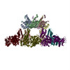
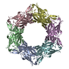
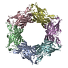
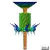
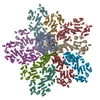
| タイトル | The X-ray structure of the gp15 hexamer and the model of the gp18 protein fitted into the cryo-EM reconstruction of the contracted T4 tail | ||||||
 要素 要素 |
| ||||||
 キーワード キーワード | VIRAL PROTEIN / bacteriophage T4 / phage tail terminator protein / phage sheath protein | ||||||
| 機能・相同性 |  機能・相同性情報 機能・相同性情報virus tail, sheath / symbiont genome ejection through host cell envelope, contractile tail mechanism / virion component 類似検索 - 分子機能 | ||||||
| 生物種 |  Enterobacteria phage T4 (ファージ) Enterobacteria phage T4 (ファージ) | ||||||
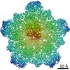
| 手法 | 電子顕微鏡法 / 単粒子再構成法 / クライオ電子顕微鏡法 / 解像度: 16 Å | ||||||
 データ登録者 データ登録者 | Fokine, A. / Zhang, Z. / Kanamaru, S. / Bowman, V.D. / Aksyuk, A. / Arisaka, F. / Rao, V.B. / Rossmann, M.G. | ||||||
 引用 引用 | ジャーナル: J Mol Biol / 年: 2013 タイトル: The molecular architecture of the bacteriophage T4 neck. 著者: Andrei Fokine / Zhihong Zhang / Shuji Kanamaru / Valorie D Bowman / Anastasia A Aksyuk / Fumio Arisaka / Venigalla B Rao / Michael G Rossmann /  要旨: A hexamer of the bacteriophage T4 tail terminator protein, gp15, attaches to the top of the phage tail stabilizing the contractile sheath and forming the interface for binding of the independently ...A hexamer of the bacteriophage T4 tail terminator protein, gp15, attaches to the top of the phage tail stabilizing the contractile sheath and forming the interface for binding of the independently assembled head. Here we report the crystal structure of the gp15 hexamer, describe its interactions in T4 virions that have either an extended tail or a contracted tail, and discuss its structural relationship to other phage proteins. The neck of T4 virions is decorated by the "collar" and "whiskers", made of fibritin molecules. Fibritin acts as a chaperone helping to attach the long tail fibers to the virus during the assembly process. The collar and whiskers are environment-sensing devices, regulating the retraction of the long tail fibers under unfavorable conditions, thus preventing infection. Cryo-electron microscopy analysis suggests that twelve fibritin molecules attach to the phage neck with six molecules forming the collar and six molecules forming the whiskers. #1: ジャーナル: Cell / 年: 2004タイトル: Three-dimensional rearrangement of proteins in the tail of bacteriophage T4 on infection of its host. 著者: Petr G Leiman / Paul R Chipman / Victor A Kostyuchenko / Vadim V Mesyanzhinov / Michael G Rossmann / 要旨: The contractile tail of bacteriophage T4 undergoes major structural transitions when the virus attaches to the host cell surface. The baseplate at the distal end of the tail changes from a hexagonal ...The contractile tail of bacteriophage T4 undergoes major structural transitions when the virus attaches to the host cell surface. The baseplate at the distal end of the tail changes from a hexagonal to a star shape. This causes the sheath around the tail tube to contract and the tail tube to protrude from the baseplate and pierce the outer cell membrane and the cell wall before reaching the inner cell membrane for subsequent viral DNA injection. Analogously, the T4 tail can be contracted by treatment with 3 M urea. The structure of the T4 contracted tail, including the head-tail joining region, has been determined by cryo-electron microscopy to 17 A resolution. This 1200 A-long, 20 MDa structure has been interpreted in terms of multiple copies of its approximately 20 component proteins. A comparison with the metastable hexagonal baseplate of the mature virus shows that the baseplate proteins move as rigid bodies relative to each other during the structural change. | ||||||
| 履歴 |
|
- 構造の表示
構造の表示
| ムービー |
ムービービューア |
|---|---|
| 構造ビューア | 分子: MolmilJmol/JSmol |
- ダウンロードとリンク
-ダウンロード
| PDBx/mmCIF形式 | 3j2n.cif.gz | 915 KB | 表示 | PDBx/mmCIF形式 |
|---|---|---|---|---|
| PDB形式 | pdb3j2n.ent.gz | 760.4 KB | 表示 | PDB形式 |
| PDBx/mmJSON形式 | 3j2n.json.gz | ツリー表示 | PDBx/mmJSON形式 | |
| その他 |  その他のダウンロード その他のダウンロード |
-検証レポート
| 文書・要旨 | 3j2n_validation.pdf.gz | 1010.5 KB | 表示 | wwPDB検証レポート |
|---|---|---|---|---|
| 文書・詳細版 | 3j2n_full_validation.pdf.gz | 1.3 MB | 表示 | |
| XML形式データ | 3j2n_validation.xml.gz | 164.6 KB | 表示 | |
| CIF形式データ | 3j2n_validation.cif.gz | 244.4 KB | 表示 | |
| アーカイブディレクトリ | https://data.pdbj.org/pub/pdb/validation_reports/j2/3j2nftp://data.pdbj.org/pub/pdb/validation_reports/j2/3j2n | HTTPS FTP |
-関連構造データ


-リンク
 PDBj
PDBj- 集合体
集合体
| 登録構造単位 | 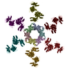
|
|---|---|
| 1 |
|
-要素
| #1: タンパク質 | 分子量: 31587.486 Da / 分子数: 6 / 由来タイプ: 組換発現 由来: (組換発現) Enterobacteria phage T4 (ファージ)遺伝子: 15 / 発現宿主:  #2: タンパク質 | 分子量: 71289.484 Da / 分子数: 6 / Mutation: R510P / 由来タイプ: 組換発現 由来: (組換発現) Enterobacteria phage T4 (ファージ)遺伝子: 18 / 発現宿主: 配列の詳細 | THE AUTHORS STATE THAT D100E, G148A, N150I, Y151I, E301G, A399V, AND H454Y ARE NATURAL VARIANTS AS ...THE AUTHORS STATE THAT D100E, G148A, N150I, Y151I, E301G, A399V, AND H454Y ARE NATURAL VARIANTS AS PER PDB ENTRY 3FOA. | |
|---|
-実験情報
-実験
| 実験 | 手法: 電子顕微鏡法 |
|---|---|
| EM実験 | 試料の集合状態: PARTICLE / 3次元再構成法: 単粒子再構成法 |
- 試料調製
試料調製
| 構成要素 |
| ||||||||||||||||||||
|---|---|---|---|---|---|---|---|---|---|---|---|---|---|---|---|---|---|---|---|---|---|
| 緩衝液 | 名称: 3 M urea / pH: 7.5 / 詳細: 3 M urea | ||||||||||||||||||||
| 試料 | 濃度: 5 mg/ml / 包埋: NO / シャドウイング: NO / 染色: NO / 凍結: YES | ||||||||||||||||||||
| 試料支持 | 詳細: 200 mesh copper grid | ||||||||||||||||||||
| 急速凍結 | 凍結剤: ETHANE / Temp: 100 K |
- 電子顕微鏡撮影
| 顕微鏡 | モデル: FEI/PHILIPS CM300FEG/T / 日付: 2002年1月6日 |
|---|---|
| 電子銃 | 電子線源:  FIELD EMISSION GUN / 加速電圧: 300 kV / 照射モード: SPOT SCAN FIELD EMISSION GUN / 加速電圧: 300 kV / 照射モード: SPOT SCAN |
| 電子レンズ | モード: BRIGHT FIELD / 倍率(公称値): 45000 X / 倍率(補正後): 47000 X / 最大 デフォーカス(公称値): 3400 nm / 最小 デフォーカス(公称値): 500 nm |
| 試料ホルダ | 試料ホルダーモデル: GATAN LIQUID NITROGEN 資料ホルダタイプ: Side entry liquid nitrogen-cooled cryo specimen holder 温度: 100 K / 傾斜角・最大: 0 ° / 傾斜角・最小: 0 ° |
| 撮影 | 電子線照射量: 20 e/Å2 / フィルム・検出器のモデル: KODAK SO-163 FILM / 詳細: Kodak SO163 film |
| 電子光学装置 | エネルギーフィルター名称: FEI |
| 画像スキャン | デジタル画像の数: 100 |
| 放射 | プロトコル: SINGLE WAVELENGTH / 単色(M)・ラウエ(L): M / 散乱光タイプ: x-ray |
| 放射波長 | 相対比: 1 |
- 解析
解析
| EMソフトウェア |
| ||||||||||||
|---|---|---|---|---|---|---|---|---|---|---|---|---|---|
| CTF補正 | 詳細: Each particle | ||||||||||||
| 対称性 | 点対称性: C6 (6回回転対称) | ||||||||||||
| 3次元再構成 | 解像度: 16 Å / 解像度の算出法: FSC 0.5 CUT-OFF / 粒子像の数: 1965 / ピクセルサイズ(公称値): 3.93 Å / ピクセルサイズ(実測値): 3.93 Å / 対称性のタイプ: POINT | ||||||||||||
| 原子モデル構築 | 空間: REAL | ||||||||||||
| 精密化ステップ | サイクル: LAST
|
