[English] 日本語
 Yorodumi
- PDB-3ezk: Bacteriophage T4 gp17 motor assembly based on crystal structures ... -
+ Open data
- Basic information
| Entry | Database: PDB / ID: 3ezk | ||||||
|---|---|---|---|---|---|---|---|
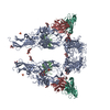
| Title | Bacteriophage T4 gp17 motor assembly based on crystal structures and cryo-EM reconstructions | ||||||
 Components Components | DNA packaging protein Gp17 | ||||||
 Keywords Keywords | HYDROLASE / pentameric motor / DNA packaging / Alternative initiation / ATP-binding / DNA-binding / Nuclease / Nucleotide-binding | ||||||
| Function / homology |  Function and homology information Function and homology informationviral terminase, large subunit / DNA nuclease activity / viral genome packaging / viral procapsid maturation / viral DNA genome packaging / nuclease activity / Hydrolases; Acting on ester bonds; Endodeoxyribonucleases producing 5'-phosphomonoesters / chromosome organization / Hydrolases; Acting on acid anhydrides; Acting on acid anhydrides to facilitate cellular and subcellular movement / endonuclease activity ...viral terminase, large subunit / DNA nuclease activity / viral genome packaging / viral procapsid maturation / viral DNA genome packaging / nuclease activity / Hydrolases; Acting on ester bonds; Endodeoxyribonucleases producing 5'-phosphomonoesters / chromosome organization / Hydrolases; Acting on acid anhydrides; Acting on acid anhydrides to facilitate cellular and subcellular movement / endonuclease activity / ATP hydrolysis activity / ATP binding / metal ion binding Similarity search - Function | ||||||
| Biological species |  Bacteriophage T4 (virus) Bacteriophage T4 (virus) | ||||||
| Method | ELECTRON MICROSCOPY / single particle reconstruction / cryo EM / Resolution: 34 Å | ||||||
 Authors Authors | Sun, S. / Rossmann, M.G. | ||||||
 Citation Citation |  Journal: Cell / Year: 2008 Journal: Cell / Year: 2008Title: The structure of the phage T4 DNA packaging motor suggests a mechanism dependent on electrostatic forces. Authors: Siyang Sun / Kiran Kondabagil / Bonnie Draper / Tanfis I Alam / Valorie D Bowman / Zhihong Zhang / Shylaja Hegde / Andrei Fokine / Michael G Rossmann / Venigalla B Rao /  Abstract: Viral genomes are packaged into "procapsids" by powerful molecular motors. We report the crystal structure of the DNA packaging motor protein, gene product 17 (gp17), in bacteriophage T4. The ...Viral genomes are packaged into "procapsids" by powerful molecular motors. We report the crystal structure of the DNA packaging motor protein, gene product 17 (gp17), in bacteriophage T4. The structure consists of an N-terminal ATPase domain, which provides energy for compacting DNA, and a C-terminal nuclease domain, which terminates packaging. We show that another function of the C-terminal domain is to translocate the genome into the procapsid. The two domains are in close contact in the crystal structure, representing a "tensed state." A cryo-electron microscopy reconstruction of the T4 procapsid complexed with gp17 shows that the packaging motor is a pentamer and that the domains within each monomer are spatially separated, representing a "relaxed state." These structures suggest a mechanism, supported by mutational and other data, in which electrostatic forces drive the DNA packaging by alternating between tensed and relaxed states. Similar mechanisms may occur in other molecular motors. | ||||||
| History |
|
- Structure visualization
| Movie |
 Movie viewer Movie viewer |
|---|---|
| Structure viewer | Molecule:  Molmil Molmil Jmol/JSmol Jmol/JSmol |
- Downloads & links
- Download
| PDBx/mmCIF format |  3ezk.cif.gz 3ezk.cif.gz | 100.9 KB | Display |  PDBx/mmCIF format PDBx/mmCIF format |
|---|---|---|---|---|
| PDB format |  pdb3ezk.ent.gz pdb3ezk.ent.gz | 62 KB | Display |  PDB format PDB format |
| PDBx/mmJSON format |  3ezk.json.gz 3ezk.json.gz | Tree view |  PDBx/mmJSON format PDBx/mmJSON format | |
| Others |  Other downloads Other downloads |
-Validation report
| Summary document |  3ezk_validation.pdf.gz 3ezk_validation.pdf.gz | 848.7 KB | Display |  wwPDB validaton report wwPDB validaton report |
|---|---|---|---|---|
| Full document |  3ezk_full_validation.pdf.gz 3ezk_full_validation.pdf.gz | 852 KB | Display | |
| Data in XML |  3ezk_validation.xml.gz 3ezk_validation.xml.gz | 36.3 KB | Display | |
| Data in CIF |  3ezk_validation.cif.gz 3ezk_validation.cif.gz | 53.7 KB | Display | |
| Arichive directory |  https://data.pdbj.org/pub/pdb/validation_reports/ez/3ezk https://data.pdbj.org/pub/pdb/validation_reports/ez/3ezk ftp://data.pdbj.org/pub/pdb/validation_reports/ez/3ezk ftp://data.pdbj.org/pub/pdb/validation_reports/ez/3ezk | HTTPS FTP |
-Related structure data
| Related structure data |  1572MC  1573C 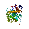 3c6aC 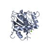 3c6hC 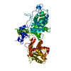 3cpeC M: map data used to model this data C: citing same article ( |
|---|---|
| Similar structure data |
- Links
Links
- Assembly
| Deposited unit | 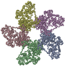
|
|---|---|
| 1 |
|
- Components
| #1: Protein | Mass: 66204.062 Da / Num. of mol.: 5 / Fragment: Residues 1-577 Source method: isolated from a genetically manipulated source Source: (gene. exp.)  Bacteriophage T4 (virus) / Strain: D / Gene: 17 / Plasmid: pET / Production host: Bacteriophage T4 (virus) / Strain: D / Gene: 17 / Plasmid: pET / Production host:  |
|---|
-Experimental details
-Experiment
| Experiment | Method: ELECTRON MICROSCOPY |
|---|---|
| EM experiment | Aggregation state: PARTICLE / 3D reconstruction method: single particle reconstruction |
- Sample preparation
| Component | Name: T4 procapsid with gp17 bound / Type: VIRUS |
|---|---|
| Buffer solution | Name: 50mM Tris-HCl, 100mM NaCl, 5mM MgCl2, 3mM beta-Mercaptoethanol pH: 7.4 Details: 50mM Tris-HCl, 100mM NaCl, 5mM MgCl2, 3mM beta-Mercaptoethanol |
| Specimen | Embedding applied: NO / Shadowing applied: NO / Staining applied: NO / Vitrification applied: YES |
| Vitrification | Instrument: HOMEMADE PLUNGER / Cryogen name: ETHANE / Details: flash-frozen on holey grids in liquid ethane |
- Electron microscopy imaging
| Microscopy | Model: FEI/PHILIPS CM200FEG |
|---|---|
| Electron gun | Electron source:  FIELD EMISSION GUN / Accelerating voltage: 200 kV / Illumination mode: FLOOD BEAM FIELD EMISSION GUN / Accelerating voltage: 200 kV / Illumination mode: FLOOD BEAM |
| Electron lens | Mode: BRIGHT FIELD / Nominal magnification: 38000 X / Nominal defocus max: 3500 nm / Nominal defocus min: 2000 nm |
| Image recording | Electron dose: 20 e/Å2 / Film or detector model: KODAK SO-163 FILM |
- Processing
| EM software |
| ||||||||||||
|---|---|---|---|---|---|---|---|---|---|---|---|---|---|
| CTF correction | Details: phase flipping of each micrograph | ||||||||||||
| Symmetry | Point symmetry: C5 (5 fold cyclic) | ||||||||||||
| 3D reconstruction | Method: Spider / Resolution: 34 Å / Num. of particles: 1716 / Nominal pixel size: 6.48 Å / Actual pixel size: 6.48 Å / Symmetry type: POINT | ||||||||||||
| Atomic model building | Protocol: RIGID BODY FIT / Space: REAL / Target criteria: Sumf Details: METHOD--N- and C-terminal domains were separately fitted into their corresponding cryoEM densities REFINEMENT PROTOCOL--Rigid body | ||||||||||||
| Atomic model building | PDB-ID: 3CPE Accession code: 3CPE / Source name: PDB / Type: experimental model | ||||||||||||
| Refinement step | Cycle: LAST
|
 Movie
Movie Controller




 PDBj
PDBj