 Movie
Movie Controller

[English] 日本語
 Yorodumi
- PDB-3psy: Endothiapepsin in complex with an inhibitor based on the Gewald r... -
+ Open data

- Basic information
| Entry | Database: PDB / ID: 3psy | ||||||
|---|---|---|---|---|---|---|---|
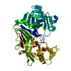
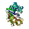
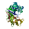
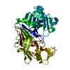
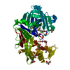
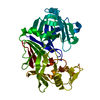
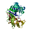
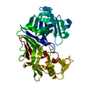
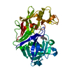
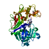
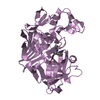
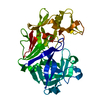
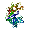
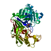
| Title | Endothiapepsin in complex with an inhibitor based on the Gewald reaction | ||||||
 Components Components | Endothiapepsin | ||||||
 Keywords Keywords | HYDROLASE/HYDROLASE INHIBITOR / HYDROLASE / HYDROLASE-HYDROLASE INHIBITOR complex | ||||||
| Function / homology |  Function and homology information Function and homology information4-hydroxy-3-polyprenylbenzoate decarboxylase / 3-octaprenyl-4-hydroxybenzoate carboxy-lyase activity / ferulate metabolic process / cinnamic acid catabolic process / ubiquinone biosynthetic process / metal ion binding / plasma membrane / cytosol Similarity search - Function | ||||||
| Biological species |  Cryphonectria parasitica (chestnut blight fungus) Cryphonectria parasitica (chestnut blight fungus) | ||||||
| Method |  X-RAY DIFFRACTION / SYNCHROTRON / MOLECULAR REPLACEMENT / Resolution: 1.43 Å X-RAY DIFFRACTION / SYNCHROTRON / MOLECULAR REPLACEMENT / Resolution: 1.43 Å | ||||||
 Authors Authors | Koester, H. / Heine, A. / Klebe, G. | ||||||
 Citation Citation | Journal: Angew.Chem.Int.Ed.Engl. / Year: 2015 Title: Tracing binding modes in hit-to-lead optimization: chameleon-like poses of aspartic protease inhibitors. Authors: Kuhnert, M. / Koster, H. / Bartholomaus, R. / Park, A.Y. / Shahim, A. / Heine, A. / Steuber, H. / Klebe, G. / Diederich, W.E. | ||||||
| History |
|
- Structure visualization
| Structure viewer | Molecule: MolmilJmol/JSmol |
|---|
- Downloads & links
-Download
| PDBx/mmCIF format | 3psy.cif.gz | 145.7 KB | Display | PDBx/mmCIF format |
|---|---|---|---|---|
| PDB format | pdb3psy.ent.gz | 111.6 KB | Display | PDB format |
| PDBx/mmJSON format | 3psy.json.gz | Tree view | PDBx/mmJSON format | |
| Others |  Other downloads Other downloads |
-Validation report
| Summary document | 3psy_validation.pdf.gz | 759.1 KB | Display | wwPDB validaton report |
|---|---|---|---|---|
| Full document | 3psy_full_validation.pdf.gz | 760.7 KB | Display | |
| Data in XML | 3psy_validation.xml.gz | 15.8 KB | Display | |
| Data in CIF | 3psy_validation.cif.gz | 23.8 KB | Display | |
| Arichive directory | https://data.pdbj.org/pub/pdb/validation_reports/ps/3psyftp://data.pdbj.org/pub/pdb/validation_reports/ps/3psy | HTTPS FTP |
-Related structure data
| Related structure data | 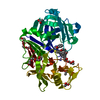 3wz6C 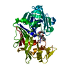 3wz7C 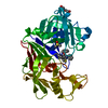 3wz8C  1oewS S: Starting model for refinement C: citing same article ( |
|---|---|
| Similar structure data |


-Links
 PDBj
PDBj
- Assembly
| Deposited unit | 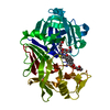
| ||||||||
|---|---|---|---|---|---|---|---|---|---|
| 1 |
| ||||||||
| Unit cell |
|
-Components
-Protein , 1 types, 1 molecules A
| #1: Protein | Mass: 33813.855 Da / Num. of mol.: 1 / Source method: isolated from a natural source Source: (natural) Cryphonectria parasitica (chestnut blight fungus)References: UniProt: P11838, EC: 3.4.23.22 |
|---|
-Non-polymers , 6 types, 240 molecules 


| #2: Chemical | ChemComp-RB9 /  Mass: 508.634 Da / Num. of mol.: 1 / Source method: obtained synthetically / Formula: C30H28N4O2S Mass: 508.634 Da / Num. of mol.: 1 / Source method: obtained synthetically / Formula: C30H28N4O2S |
|---|---|
| #3: Chemical | ChemComp-DMS /  Mass: 78.133 Da / Num. of mol.: 1 / Source method: obtained synthetically / Formula: C2H6OS / Comment: DMSO, precipitant*YM Mass: 78.133 Da / Num. of mol.: 1 / Source method: obtained synthetically / Formula: C2H6OS / Comment: DMSO, precipitant*YM |
| #4: Chemical | ChemComp-GOL /  Mass: 92.094 Da / Num. of mol.: 1 / Source method: obtained synthetically / Formula: C3H8O3 Mass: 92.094 Da / Num. of mol.: 1 / Source method: obtained synthetically / Formula: C3H8O3 |
| #5: Chemical | ChemComp-PG4 /  Mass: 194.226 Da / Num. of mol.: 1 / Source method: obtained synthetically / Formula: C8H18O5 / Comment: precipitant*YM Mass: 194.226 Da / Num. of mol.: 1 / Source method: obtained synthetically / Formula: C8H18O5 / Comment: precipitant*YM |
| #6: Chemical | ChemComp-1PE /  Mass: 238.278 Da / Num. of mol.: 1 / Source method: obtained synthetically / Formula: C10H22O6 / Comment: precipitant*YM Mass: 238.278 Da / Num. of mol.: 1 / Source method: obtained synthetically / Formula: C10H22O6 / Comment: precipitant*YM |
| #7: Water | ChemComp-HOH / Mass: 18.015 Da / Num. of mol.: 235 / Source method: isolated from a natural source / Formula: H2O |
-Experimental details
-Experiment
| Experiment | Method: X-RAY DIFFRACTION / Number of used crystals: 1 |
|---|
- Sample preparation
| Crystal | Density Matthews: 2.44 Å3/Da / Density % sol: 49.64 % |
|---|---|
| Crystal grow | Temperature: 289 K / Method: vapor diffusion, sitting drop / pH: 4.6 Details: 0.1M NH4Ac, 0.1M Acetate-Buffer pH 4.6, 26% PEG 4000, VAPOR DIFFUSION, SITTING DROP, temperature 289K |
-Data collection
| Diffraction | Mean temperature: 100 K |
|---|---|
| Diffraction source | Source: SYNCHROTRON / Site: SLS  / Beamline: X06DA / Wavelength: 1 Å / Beamline: X06DA / Wavelength: 1 Å |
| Detector | Type: MARMOSAIC 225 mm CCD / Detector: CCD / Date: Jun 22, 2009 / Details: mirrors |
| Radiation | Monochromator: Bartels Monochromator with dual channel cut crystals (DCCM) in (+--+) geometry Protocol: SINGLE WAVELENGTH / Monochromatic (M) / Laue (L): M / Scattering type: x-ray |
| Radiation wavelength | Wavelength: 1 Å / Relative weight: 1 |
| Reflection | Resolution: 1.43→40 Å / Num. all: 55570 / Num. obs: 55570 / % possible obs: 100 % / Redundancy: 3.5 % / Rsym value: 0.034 / Net I/σ(I): 34.2 |
| Reflection shell | Resolution: 1.43→1.45 Å / Redundancy: 3.3 % / Mean I/σ(I) obs: 4.2 / Num. unique all: 2578 / Rsym value: 0.285 / % possible all: 100 |
- Processing
| Software |
| |||||||||||||||||||||||||||||||||
|---|---|---|---|---|---|---|---|---|---|---|---|---|---|---|---|---|---|---|---|---|---|---|---|---|---|---|---|---|---|---|---|---|---|---|
| Refinement | Method to determine structure: MOLECULAR REPLACEMENT Starting model: 1OEW Resolution: 1.43→10 Å / Num. parameters: 24349 / Num. restraintsaints: 30620 / Cross valid method: THROUGHOUT / σ(F): 0 / Stereochemistry target values: Engh & Huber
| |||||||||||||||||||||||||||||||||
| Refine analyze | Num. disordered residues: 6 / Occupancy sum hydrogen: 2239.64 / Occupancy sum non hydrogen: 2676.32 | |||||||||||||||||||||||||||||||||
| Refinement step | Cycle: LAST / Resolution: 1.43→10 Å
| |||||||||||||||||||||||||||||||||
| Refine LS restraints |
|
