 Movie
Movie Controller

[English] 日本語
 Yorodumi
- PDB-3oig: Crystal Structure of Enoyl-ACP Reductases I (FabI) from B. subtil... -
+ Open data

- Basic information
| Entry | Database: PDB / ID: 3oig | ||||||
|---|---|---|---|---|---|---|---|
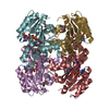
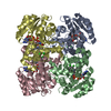
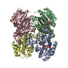
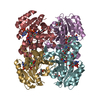
| Title | Crystal Structure of Enoyl-ACP Reductases I (FabI) from B. subtilis (complex with NAD and INH) | ||||||
 Components Components | Enoyl-[acyl-carrier-protein] reductase [NADH] | ||||||
 Keywords Keywords | OXIDOREDUCTASE/OXIDOREDUCTASE inhibitor / Fatty acid synthesis / Rossmann-like fold / Enoyl-ACP Reductases / NADH binding / OXIDOREDUCTASE-OXIDOREDUCTASE inhibitor complex | ||||||
| Function / homology |  Function and homology information Function and homology informationenoyl-[acyl-carrier-protein] reductase (NADH) / fatty acid elongation / enoyl-[acyl-carrier-protein] reductase (NADH) activity / cellular response to cold Similarity search - Function | ||||||
| Biological species |  | ||||||
| Method |  X-RAY DIFFRACTION / SYNCHROTRON / MOLECULAR REPLACEMENT / Resolution: 1.25 Å X-RAY DIFFRACTION / SYNCHROTRON / MOLECULAR REPLACEMENT / Resolution: 1.25 Å | ||||||
 Authors Authors | Kim, K.-H. / Ha, B.H. / Kim, S.J. / Hong, S.K. / Hwang, K.Y. / Kim, E.E. | ||||||
 Citation Citation | Journal: J.Mol.Biol. / Year: 2011 Title: Crystal Structures of Enoyl-ACP Reductases I (FabI) and III (FabL) from B. subtilis Authors: Kim, K.-H. / Ha, B.H. / Kim, S.J. / Hong, S.K. / Hwang, K.Y. / Kim, E.E. | ||||||
| History |
|
- Structure visualization
| Structure viewer | Molecule: MolmilJmol/JSmol |
|---|
- Downloads & links
-Download
| PDBx/mmCIF format | 3oig.cif.gz | 73.5 KB | Display | PDBx/mmCIF format |
|---|---|---|---|---|
| PDB format | pdb3oig.ent.gz | 52.8 KB | Display | PDB format |
| PDBx/mmJSON format | 3oig.json.gz | Tree view | PDBx/mmJSON format | |
| Others |  Other downloads Other downloads |
-Validation report
| Arichive directory | https://data.pdbj.org/pub/pdb/validation_reports/oi/3oigftp://data.pdbj.org/pub/pdb/validation_reports/oi/3oig | HTTPS FTP |
|---|
-Related structure data
| Related structure data |  3oicC  3oidC  3oifSC C: citing same article ( S: Starting model for refinement |
|---|---|
| Similar structure data |


-Links
 PDBj
PDBj

- Assembly
| Deposited unit | 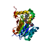
| ||||||||
|---|---|---|---|---|---|---|---|---|---|
| 1 | 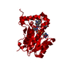
| ||||||||
| Unit cell |
| ||||||||
| Components on special symmetry positions |
|
-Components
| #1: Protein | Mass: 28976.879 Da / Num. of mol.: 1 Source method: isolated from a genetically manipulated source Source: (gene. exp.) References: UniProt: P54616, enoyl-[acyl-carrier-protein] reductase (NADH) |
|---|---|
| #2: Chemical | ChemComp-NAD /   Mass: 663.425 Da / Num. of mol.: 1 / Source method: obtained synthetically / Formula: C21H27N7O14P2 / Comment: NAD*YM Mass: 663.425 Da / Num. of mol.: 1 / Source method: obtained synthetically / Formula: C21H27N7O14P2 / Comment: NAD*YM |
| #3: Chemical | ChemComp-IMJ / (  Mass: 388.462 Da / Num. of mol.: 1 / Source method: obtained synthetically / Formula: C23H24N4O2 Mass: 388.462 Da / Num. of mol.: 1 / Source method: obtained synthetically / Formula: C23H24N4O2 |
| #4: Water | ChemComp-HOH /  Mass: 18.015 Da / Num. of mol.: 265 / Source method: isolated from a natural source / Formula: H2O Mass: 18.015 Da / Num. of mol.: 265 / Source method: isolated from a natural source / Formula: H2O |
-Experimental details
-Experiment
| Experiment | Method: X-RAY DIFFRACTION / Number of used crystals: 1 |
|---|
- Sample preparation
| Crystal | Density Matthews: 2.1 Å3/Da / Density % sol: 41.46 % |
|---|---|
| Crystal grow | Temperature: 277 K / Method: vapor diffusion, hanging drop / pH: 8 Details: 0.2M di-ammonium tartrate, 22% (w/v) PEG 3350, pH 8.0, VAPOR DIFFUSION, HANGING DROP, temperature 277K |
-Data collection
| Diffraction | Mean temperature: 100 K |
|---|---|
| Diffraction source | Source: SYNCHROTRON / Site: PAL/PLS  / Beamline: 6B / Wavelength: 1.12174 Å / Beamline: 6B / Wavelength: 1.12174 Å |
| Detector | Type: Bruker Proteum 300 / Detector: CCD / Date: Nov 15, 2005 / Details: mirrors |
| Radiation | Protocol: SINGLE WAVELENGTH / Monochromatic (M) / Laue (L): M / Scattering type: x-ray |
| Radiation wavelength | Wavelength: 1.12174 Å / Relative weight: 1 |
| Reflection | Resolution: 1.25→50 Å / Num. obs: 67984 / % possible obs: 99.8 % / Observed criterion σ(F): 1 / Observed criterion σ(I): 1 / Redundancy: 3.1 % / Rmerge(I) obs: 0.099 / Net I/σ(I): 19.8 / Num. measured all: 672557 |
| Reflection shell | Resolution: 1.25→1.29 Å / Redundancy: 2.8 % / Rmerge(I) obs: 0.47 / Mean I/σ(I) obs: 1.93 |
- Processing
| Software |
| |||||||||||||||||||||||||||||||||||||||||||||||||||||||||||||||||
|---|---|---|---|---|---|---|---|---|---|---|---|---|---|---|---|---|---|---|---|---|---|---|---|---|---|---|---|---|---|---|---|---|---|---|---|---|---|---|---|---|---|---|---|---|---|---|---|---|---|---|---|---|---|---|---|---|---|---|---|---|---|---|---|---|---|---|
| Refinement | Method to determine structure: MOLECULAR REPLACEMENT Starting model: PDB ENTRY 3OIF Resolution: 1.25→33.69 Å / Cor.coef. Fo:Fc: 0.964 / Cor.coef. Fo:Fc free: 0.955 / SU B: 0.56 / SU ML: 0.025 / Cross valid method: THROUGHOUT / σ(F): 0 / ESU R Free: 0.045 / Stereochemistry target values: MAXIMUM LIKELIHOOD / Details: HYDROGENS HAVE BEEN ADDED IN THE RIDING POSITIONS
| |||||||||||||||||||||||||||||||||||||||||||||||||||||||||||||||||
| Solvent computation | Ion probe radii: 0.8 Å / Shrinkage radii: 0.8 Å / VDW probe radii: 1.4 Å / Solvent model: MASK | |||||||||||||||||||||||||||||||||||||||||||||||||||||||||||||||||
| Displacement parameters | Biso mean: 11.324 Å2
| |||||||||||||||||||||||||||||||||||||||||||||||||||||||||||||||||
| Refinement step | Cycle: LAST / Resolution: 1.25→33.69 Å
| |||||||||||||||||||||||||||||||||||||||||||||||||||||||||||||||||
| Refine LS restraints |
| |||||||||||||||||||||||||||||||||||||||||||||||||||||||||||||||||
| LS refinement shell | Resolution: 1.247→1.28 Å / Total num. of bins used: 20
|
