 Movie
Movie Controller

[English] 日本語
 Yorodumi
- PDB-3oif: Crystal Structure of Enoyl-ACP Reductases I (FabI) from B. subtil... -
+ Open data

- Basic information
| Entry | Database: PDB / ID: 3oif | ||||||
|---|---|---|---|---|---|---|---|
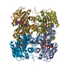
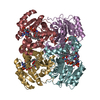
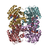
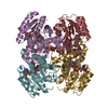
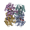
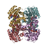
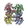
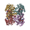
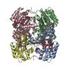
| Title | Crystal Structure of Enoyl-ACP Reductases I (FabI) from B. subtilis (complex with NAD and TCL) | ||||||
 Components Components | Enoyl-[acyl-carrier-protein] reductase [NADH] | ||||||
 Keywords Keywords | OXIDOREDUCTASE / Fatty acid synthesis / Rossmann-like fold / Enoyl-ACP Reductases / NADH binding | ||||||
| Function / homology |  Function and homology information Function and homology informationenoyl-[acyl-carrier-protein] reductase (NADH) / fatty acid elongation / enoyl-[acyl-carrier-protein] reductase (NADH) activity / cellular response to cold Similarity search - Function | ||||||
| Biological species |  | ||||||
| Method |  X-RAY DIFFRACTION / SYNCHROTRON / MOLECULAR REPLACEMENT / Resolution: 2.6 Å X-RAY DIFFRACTION / SYNCHROTRON / MOLECULAR REPLACEMENT / Resolution: 2.6 Å | ||||||
 Authors Authors | Kim, K.-H. / Ha, B.H. / Kim, S.J. / Hong, S.K. / Hwang, K.Y. / Kim, E.E. | ||||||
 Citation Citation | Journal: J.Mol.Biol. / Year: 2011 Title: Crystal Structures of Enoyl-ACP Reductases I (FabI) and III (FabL) from B. subtilis Authors: Kim, K.-H. / Ha, B.H. / Kim, S.J. / Hong, S.K. / Hwang, K.Y. / Kim, E.E. | ||||||
| History |
|
- Structure visualization
| Structure viewer | Molecule: MolmilJmol/JSmol |
|---|
- Downloads & links
-Download
| PDBx/mmCIF format | 3oif.cif.gz | 196.9 KB | Display | PDBx/mmCIF format |
|---|---|---|---|---|
| PDB format | pdb3oif.ent.gz | 159.1 KB | Display | PDB format |
| PDBx/mmJSON format | 3oif.json.gz | Tree view | PDBx/mmJSON format | |
| Others |  Other downloads Other downloads |
-Validation report
| Arichive directory | https://data.pdbj.org/pub/pdb/validation_reports/oi/3oifftp://data.pdbj.org/pub/pdb/validation_reports/oi/3oif | HTTPS FTP |
|---|
-Related structure data
| Related structure data |  3oicC  3oidC 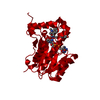 3oigC 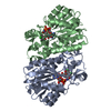 1c14S C: citing same article ( S: Starting model for refinement |
|---|---|
| Similar structure data |

-Links
 PDBj
PDBj

- Assembly
| Deposited unit | 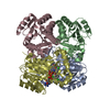
| ||||||||
|---|---|---|---|---|---|---|---|---|---|
| 1 |
| ||||||||
| Unit cell |
|
-Components
| #1: Protein | Mass: 28976.879 Da / Num. of mol.: 4 Source method: isolated from a genetically manipulated source Source: (gene. exp.) References: UniProt: P54616, enoyl-[acyl-carrier-protein] reductase (NADH) #2: Chemical |   Mass: 663.425 Da / Num. of mol.: 2 / Source method: obtained synthetically / Formula: C21H27N7O14P2 / Comment: NAD*YM Mass: 663.425 Da / Num. of mol.: 2 / Source method: obtained synthetically / Formula: C21H27N7O14P2 / Comment: NAD*YM#3: Chemical |   Mass: 289.542 Da / Num. of mol.: 2 / Source method: obtained synthetically / Formula: C12H7Cl3O2 / Comment: antifungal, antibiotic, detergent*YM Mass: 289.542 Da / Num. of mol.: 2 / Source method: obtained synthetically / Formula: C12H7Cl3O2 / Comment: antifungal, antibiotic, detergent*YM#4: Water | ChemComp-HOH / |  Mass: 18.015 Da / Num. of mol.: 132 / Source method: isolated from a natural source / Formula: H2O Mass: 18.015 Da / Num. of mol.: 132 / Source method: isolated from a natural source / Formula: H2O |
|---|
-Experimental details
-Experiment
| Experiment | Method: X-RAY DIFFRACTION / Number of used crystals: 1 |
|---|
- Sample preparation
| Crystal | Density Matthews: 2.35 Å3/Da / Density % sol: 47.75 % |
|---|---|
| Crystal grow | Temperature: 277 K / Method: vapor diffusion, hanging drop / pH: 8 Details: 0.2M di-ammonium tartrate, 20% (w/v) PEG 3350, pH 8.0, VAPOR DIFFUSION, HANGING DROP, temperature 277K |
-Data collection
| Diffraction | Mean temperature: 100 K |
|---|---|
| Diffraction source | Source: SYNCHROTRON / Site: PAL/PLS  / Beamline: 6B / Wavelength: 1.12174 Å / Beamline: 6B / Wavelength: 1.12174 Å |
| Detector | Type: Bruker Proteum 300 / Detector: CCD / Date: Nov 15, 2005 / Details: mirrors |
| Radiation | Protocol: SINGLE WAVELENGTH / Monochromatic (M) / Laue (L): M / Scattering type: x-ray |
| Radiation wavelength | Wavelength: 1.12174 Å / Relative weight: 1 |
| Reflection | Resolution: 2.6→30 Å / Num. obs: 34401 / % possible obs: 96 % / Observed criterion σ(F): 1 / Observed criterion σ(I): 1 / Redundancy: 2.5 % / Rmerge(I) obs: 0.108 / Net I/σ(I): 13.2 / Num. measured all: 623716 |
| Reflection shell | Resolution: 2.6→2.69 Å / Redundancy: 2.1 % / Rmerge(I) obs: 0.283 / Mean I/σ(I) obs: 1.77 / % possible all: 91.2 |
- Processing
| Software |
| |||||||||||||||||||||||||||||||||||||||||||||||||||||||||||||||||
|---|---|---|---|---|---|---|---|---|---|---|---|---|---|---|---|---|---|---|---|---|---|---|---|---|---|---|---|---|---|---|---|---|---|---|---|---|---|---|---|---|---|---|---|---|---|---|---|---|---|---|---|---|---|---|---|---|---|---|---|---|---|---|---|---|---|---|
| Refinement | Method to determine structure: MOLECULAR REPLACEMENT Starting model: PDB ENTRY 1C14 Resolution: 2.6→29.98 Å / Cor.coef. Fo:Fc: 0.935 / Cor.coef. Fo:Fc free: 0.891 / SU B: 12.664 / SU ML: 0.27 / Cross valid method: THROUGHOUT / σ(F): 0 / ESU R Free: 0.37 / Stereochemistry target values: MAXIMUM LIKELIHOOD / Details: HYDROGENS HAVE BEEN ADDED IN THE RIDING POSITIONS
| |||||||||||||||||||||||||||||||||||||||||||||||||||||||||||||||||
| Solvent computation | Ion probe radii: 0.8 Å / Shrinkage radii: 0.8 Å / VDW probe radii: 1.4 Å / Solvent model: MASK | |||||||||||||||||||||||||||||||||||||||||||||||||||||||||||||||||
| Displacement parameters | Biso mean: 49.041 Å2
| |||||||||||||||||||||||||||||||||||||||||||||||||||||||||||||||||
| Refinement step | Cycle: LAST / Resolution: 2.6→29.98 Å
| |||||||||||||||||||||||||||||||||||||||||||||||||||||||||||||||||
| Refine LS restraints |
| |||||||||||||||||||||||||||||||||||||||||||||||||||||||||||||||||
| LS refinement shell | Resolution: 2.604→2.672 Å / Total num. of bins used: 20
|
