 Movie
Movie Controller

[English] 日本語
 Yorodumi
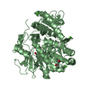
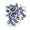
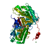
- PDB-3o82: Structure of BasE N-terminal domain from Acinetobacter baumannii ... -
+ Open data

- Basic information
| Entry | Database: PDB / ID: 3o82 | ||||||
|---|---|---|---|---|---|---|---|
| Title | Structure of BasE N-terminal domain from Acinetobacter baumannii bound to 5'-O-[N-(2,3-dihydroxybenzoyl)sulfamoyl] adenosine | ||||||
 Components Components | Peptide arylation enzyme | ||||||
 Keywords Keywords | LIGASE / Adenylation of 2 / 3-dihydroxybenzoate and transfer to pantetheine cofactor of BasF / Non-Ribosomal Peptide Synthetase (NRPS) | ||||||
| Function / homology |  Function and homology information Function and homology information2,3-dihydroxybenzoate--[aryl-carrier protein] ligase / siderophore biosynthetic process Similarity search - Function | ||||||
| Biological species |  Acinetobacter baumannii (bacteria) Acinetobacter baumannii (bacteria) | ||||||
| Method |  X-RAY DIFFRACTION / SYNCHROTRON / MOLECULAR REPLACEMENT / Resolution: 2.7 Å X-RAY DIFFRACTION / SYNCHROTRON / MOLECULAR REPLACEMENT / Resolution: 2.7 Å | ||||||
 Authors Authors | Drake, E.J. / Duckworth, B.P. / Neres, J. / Aldrich, C.C. / Gulick, A.M. | ||||||
 Citation Citation | Journal: Biochemistry / Year: 2010 Title: Biochemical and structural characterization of bisubstrate inhibitors of BasE, the self-standing nonribosomal peptide synthetase adenylate-forming enzyme of acinetobactin synthesis. Authors: Drake, E.J. / Duckworth, B.P. / Neres, J. / Aldrich, C.C. / Gulick, A.M. | ||||||
| History |
|
- Structure visualization
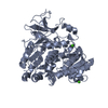
| Structure viewer | Molecule: MolmilJmol/JSmol |
|---|
- Downloads & links
-Download
| PDBx/mmCIF format | 3o82.cif.gz | 186 KB | Display | PDBx/mmCIF format |
|---|---|---|---|---|
| PDB format | pdb3o82.ent.gz | 143.5 KB | Display | PDB format |
| PDBx/mmJSON format | 3o82.json.gz | Tree view | PDBx/mmJSON format | |
| Others |  Other downloads Other downloads |
-Validation report
| Summary document | 3o82_validation.pdf.gz | 1.2 MB | Display | wwPDB validaton report |
|---|---|---|---|---|
| Full document | 3o82_full_validation.pdf.gz | 1.2 MB | Display | |
| Data in XML | 3o82_validation.xml.gz | 32.6 KB | Display | |
| Data in CIF | 3o82_validation.cif.gz | 44.8 KB | Display | |
| Arichive directory | https://data.pdbj.org/pub/pdb/validation_reports/o8/3o82ftp://data.pdbj.org/pub/pdb/validation_reports/o8/3o82 | HTTPS FTP |
-Related structure data
| Related structure data | 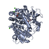 3o83C 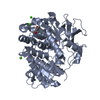 3o84C  1mdbS C: citing same article ( S: Starting model for refinement |
|---|---|
| Similar structure data |


-Links
 PDBj
PDBj
- Assembly
| Deposited unit | 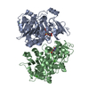
| ||||||||||||||||||
|---|---|---|---|---|---|---|---|---|---|---|---|---|---|---|---|---|---|---|---|
| 1 | 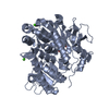
| ||||||||||||||||||
| 2 | 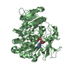
| ||||||||||||||||||
| 3 |
| ||||||||||||||||||
| Unit cell |
| ||||||||||||||||||
| Noncrystallographic symmetry (NCS) | NCS domain:
NCS domain segments: Component-ID: 1 / Ens-ID: 1 / Beg auth comp-ID: LEU / Beg label comp-ID: LEU / End auth comp-ID: ARG / End label comp-ID: ARG / Refine code: 4 / Auth seq-ID: 5 - 435 / Label seq-ID: 7 - 437
|
-Components
| #1: Protein | Mass: 60868.270 Da / Num. of mol.: 2 / Fragment: BasE / Mutation: P45L Source method: isolated from a genetically manipulated source Source: (gene. exp.) Acinetobacter baumannii (bacteria) / Strain: AB900 / Gene: ACICU_02578, basE / Plasmid: pED453 / Production host: References: UniProt: B2HVG8, UniProt: A0A7U3Y1M5*PLUS, Ligases; Forming carbon-sulfur bonds; Acid-thiol ligases #2: Chemical | 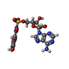  Mass: 482.425 Da / Num. of mol.: 2 / Source method: obtained synthetically / Formula: C17H18N6O9S Mass: 482.425 Da / Num. of mol.: 2 / Source method: obtained synthetically / Formula: C17H18N6O9S#3: Chemical | ChemComp-CA /   Mass: 40.078 Da / Num. of mol.: 4 / Source method: obtained synthetically / Formula: Ca Mass: 40.078 Da / Num. of mol.: 4 / Source method: obtained synthetically / Formula: Ca#4: Water | ChemComp-HOH / |  Mass: 18.015 Da / Num. of mol.: 49 / Source method: isolated from a natural source / Formula: H2O Mass: 18.015 Da / Num. of mol.: 49 / Source method: isolated from a natural source / Formula: H2OSequence details | THIS ENTRY USES A UNIPROT REFERENCE THAT IS FOR A DIFFERENT STRAIN OF A. BAUMANNI. THESE CHANGES ...THIS ENTRY USES A UNIPROT REFERENCE THAT IS FOR A DIFFERENT STRAIN OF A. BAUMANNI. THESE CHANGES ARE STRAIN RELATED DIFFERENCE | |
|---|
-Experimental details
-Experiment
| Experiment | Method: X-RAY DIFFRACTION / Number of used crystals: 1 |
|---|
- Sample preparation
| Crystal | Density Matthews: 2.91 Å3/Da / Density % sol: 57.73 % |
|---|---|
| Crystal grow | Temperature: 287 K / Method: vapor diffusion, hanging drop / pH: 7.5 Details: 5-15% PEG 8000, 5% MPD, 250-600 mM CaCl2, 50 mM BTP, pH 7.5, VAPOR DIFFUSION, HANGING DROP, temperature 287K |
-Data collection
| Diffraction | Mean temperature: 113 K |
|---|---|
| Diffraction source | Source: SYNCHROTRON / Site: SSRL  / Beamline: BL9-1 / Wavelength: 0.979 Å / Beamline: BL9-1 / Wavelength: 0.979 Å |
| Detector | Type: ADSC QUANTUM 315r / Detector: CCD / Date: Feb 25, 2008 |
| Radiation | Monochromator: Side-scattering cuberoot I-beam bent single crystal; asymetric cut 12.2 degs Protocol: SINGLE WAVELENGTH / Monochromatic (M) / Laue (L): M / Scattering type: x-ray |
| Radiation wavelength | Wavelength: 0.979 Å / Relative weight: 1 |
| Reflection | Resolution: 2.7→40 Å / Num. all: 39977 / Num. obs: 38979 / % possible obs: 97.5 % / Observed criterion σ(F): 0 / Observed criterion σ(I): -3 / Redundancy: 3.8 % / Biso Wilson estimate: 56.6 Å2 / Rsym value: 0.131 / Net I/σ(I): 9.2 |
| Reflection shell | Resolution: 2.7→2.8 Å / Redundancy: 3.2 % / Mean I/σ(I) obs: 2.2 / Rsym value: 0.377 / % possible all: 81.9 |
- Processing
| Software |
| |||||||||||||||||||||||||||||||||||||||||||||||||||||||||||||||||
|---|---|---|---|---|---|---|---|---|---|---|---|---|---|---|---|---|---|---|---|---|---|---|---|---|---|---|---|---|---|---|---|---|---|---|---|---|---|---|---|---|---|---|---|---|---|---|---|---|---|---|---|---|---|---|---|---|---|---|---|---|---|---|---|---|---|---|
| Refinement | Method to determine structure: MOLECULAR REPLACEMENT Starting model: PDB ENTRY 1MDB residues 1-430 Resolution: 2.7→40 Å / Cor.coef. Fo:Fc: 0.932 / Cor.coef. Fo:Fc free: 0.896 / SU B: 10.586 / SU ML: 0.219 / Cross valid method: THROUGHOUT / ESU R: 0.475 / ESU R Free: 0.301 / Stereochemistry target values: MAXIMUM LIKELIHOOD / Details: HYDROGENS HAVE BEEN ADDED IN THE RIDING POSITIONS
| |||||||||||||||||||||||||||||||||||||||||||||||||||||||||||||||||
| Solvent computation | Ion probe radii: 0.8 Å / Shrinkage radii: 0.8 Å / VDW probe radii: 1.4 Å / Solvent model: BABINET MODEL WITH MASK | |||||||||||||||||||||||||||||||||||||||||||||||||||||||||||||||||
| Displacement parameters | Biso mean: 57.064 Å2
| |||||||||||||||||||||||||||||||||||||||||||||||||||||||||||||||||
| Refinement step | Cycle: LAST / Resolution: 2.7→40 Å
| |||||||||||||||||||||||||||||||||||||||||||||||||||||||||||||||||
| Refine LS restraints |
| |||||||||||||||||||||||||||||||||||||||||||||||||||||||||||||||||
| Refine LS restraints NCS | Dom-ID: 1 / Auth asym-ID: A / Ens-ID: 1 / Number: 3323 / Refine-ID: X-RAY DIFFRACTION
| |||||||||||||||||||||||||||||||||||||||||||||||||||||||||||||||||
| LS refinement shell | Resolution: 2.7→2.77 Å / Total num. of bins used: 20
|
