 Movie
Movie Controller

[English] 日本語
 Yorodumi
- PDB-3nyf: Crystal Structure of Pseudomonas aeruginosa D-Arginine Dehydrogen... -
+ Open data

- Basic information
| Entry | Database: PDB / ID: 3nyf | ||||||
|---|---|---|---|---|---|---|---|
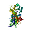
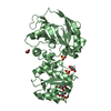
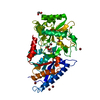
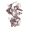
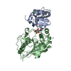
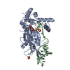
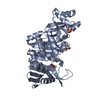
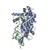
| Title | Crystal Structure of Pseudomonas aeruginosa D-Arginine Dehydrogenase in Complex with Imino-Histidine | ||||||
 Components Components | D-Arginine Dehydrogenase | ||||||
 Keywords Keywords | OXIDOREDUCTASE / D-Arginine Dehydrogenase / FAD / Imino-Histidine | ||||||
| Function / homology |  Function and homology information Function and homology informationD-arginine dehydrogenase / D-amino-acid dehydrogenase activity / arginine metabolic process / L-arginine catabolic process / nucleotide binding / cytoplasm Similarity search - Function | ||||||
| Biological species |   Pseudomonas aeruginosa (bacteria) Pseudomonas aeruginosa (bacteria) | ||||||
| Method |  X-RAY DIFFRACTION / SYNCHROTRON / MOLECULAR REPLACEMENT / Resolution: 1.3 Å X-RAY DIFFRACTION / SYNCHROTRON / MOLECULAR REPLACEMENT / Resolution: 1.3 Å | ||||||
 Authors Authors | Fu, G. / Weber, I.T. | ||||||
 Citation Citation | Journal: Biochemistry / Year: 2010 Title: Conformational changes and substrate recognition in Pseudomonas aeruginosa D-arginine dehydrogenase. Authors: Fu, G. / Yuan, H. / Li, C. / Lu, C.D. / Gadda, G. / Weber, I.T. | ||||||
| History |
|
- Structure visualization
| Structure viewer | Molecule: MolmilJmol/JSmol |
|---|
- Downloads & links
-Download
| PDBx/mmCIF format | 3nyf.cif.gz | 181.1 KB | Display | PDBx/mmCIF format |
|---|---|---|---|---|
| PDB format | pdb3nyf.ent.gz | 140.1 KB | Display | PDB format |
| PDBx/mmJSON format | 3nyf.json.gz | Tree view | PDBx/mmJSON format | |
| Others |  Other downloads Other downloads |
-Validation report
| Arichive directory | https://data.pdbj.org/pub/pdb/validation_reports/ny/3nyfftp://data.pdbj.org/pub/pdb/validation_reports/ny/3nyf | HTTPS FTP |
|---|
-Related structure data
| Related structure data |  3nycSC 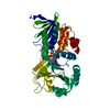 3nyeC S: Starting model for refinement C: citing same article ( |
|---|---|
| Similar structure data |


-Links
 PDBj
PDBj- Assembly
| Deposited unit | 
| ||||||||
|---|---|---|---|---|---|---|---|---|---|
| 1 |
| ||||||||
| Unit cell |
|
-Components
| #1: Protein | Mass: 41405.504 Da / Num. of mol.: 1 Source method: isolated from a genetically manipulated source Source: (gene. exp.) Pseudomonas aeruginosa (bacteria) / Strain: PAO1 / Gene: PA3863 / Plasmid: pBAD-His-6 / Production host: References: UniProt: Q9HXE3, Oxidoreductases; Acting on the CH-NH2 group of donors; With NAD+ or NADP+ as acceptor |
|---|---|
| #2: Chemical | ChemComp-FAD /   Mass: 785.550 Da / Num. of mol.: 1 / Source method: obtained synthetically / Formula: C27H33N9O15P2 / Comment: FAD*YM Mass: 785.550 Da / Num. of mol.: 1 / Source method: obtained synthetically / Formula: C27H33N9O15P2 / Comment: FAD*YM |
| #3: Chemical | ChemComp-HHI / (  Type: peptide linking / Mass: 153.139 Da / Num. of mol.: 1 / Source method: obtained synthetically / Formula: C6H7N3O2 Type: peptide linking / Mass: 153.139 Da / Num. of mol.: 1 / Source method: obtained synthetically / Formula: C6H7N3O2 |
| #4: Water | ChemComp-HOH /  Mass: 18.015 Da / Num. of mol.: 353 / Source method: isolated from a natural source / Formula: H2O Mass: 18.015 Da / Num. of mol.: 353 / Source method: isolated from a natural source / Formula: H2O |
-Experimental details
-Experiment
| Experiment | Method: X-RAY DIFFRACTION / Number of used crystals: 1 |
|---|
- Sample preparation
| Crystal | Density Matthews: 2.63 Å3/Da / Density % sol: 53.15 % |
|---|---|
| Crystal grow | Temperature: 277 K / Method: vapor diffusion, hanging drop / pH: 6.8 Details: 0.1M 2-(N-morpholino)-ethanesulfonic acid (MES) pH 6.5-7.0, 5% glycerol, 6-10% (w/v) PEG6000, VAPOR DIFFUSION, HANGING DROP, temperature 277K |
-Data collection
| Diffraction | Mean temperature: 100 K |
|---|---|
| Diffraction source | Source: SYNCHROTRON / Site: APS  / Beamline: 22-BM / Wavelength: 1 Å / Beamline: 22-BM / Wavelength: 1 Å |
| Detector | Type: MARMOSAIC 225 mm CCD / Detector: CCD / Date: Mar 27, 2009 |
| Radiation | Protocol: SINGLE WAVELENGTH / Monochromatic (M) / Laue (L): M / Scattering type: x-ray |
| Radiation wavelength | Wavelength: 1 Å / Relative weight: 1 |
| Reflection | Resolution: 1.3→50 Å / Num. all: 97110 / Num. obs: 97110 / % possible obs: 90.4 % / Observed criterion σ(F): 0 / Observed criterion σ(I): 0 / Redundancy: 6.5 % / Rsym value: 0.049 / Net I/σ(I): 3.2 |
| Reflection shell | Resolution: 1.3→1.35 Å / Redundancy: 2.7 % / Rmerge(I) obs: 0.306 / Mean I/σ(I) obs: 3.2 / Num. unique all: 5814 / % possible all: 54.7 |
- Processing
| Software |
| |||||||||||||||||||||||||||||||||
|---|---|---|---|---|---|---|---|---|---|---|---|---|---|---|---|---|---|---|---|---|---|---|---|---|---|---|---|---|---|---|---|---|---|---|
| Refinement | Method to determine structure: MOLECULAR REPLACEMENT Starting model: PDB entry 3NYC Resolution: 1.3→10 Å / Num. parameters: 30605 / Num. restraintsaints: 36537 / Cross valid method: FREE R / σ(F): 0 / Stereochemistry target values: ENGH AND HUBER / Details: conjugage gradient minimization
| |||||||||||||||||||||||||||||||||
| Refine analyze | Num. disordered residues: 11 / Occupancy sum hydrogen: 0 / Occupancy sum non hydrogen: 3330.3 | |||||||||||||||||||||||||||||||||
| Refinement step | Cycle: LAST / Resolution: 1.3→10 Å
| |||||||||||||||||||||||||||||||||
| Refine LS restraints |
|
