 Movie
Movie Controller

[English] 日本語
 Yorodumi
- PDB-3eki: Structural insights of the Mycoplasma hyorhinis protein Mh-p37: A... -
+ Open data

- Basic information
| Entry | Database: PDB / ID: 3eki | ||||||
|---|---|---|---|---|---|---|---|
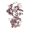
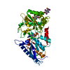
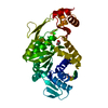
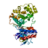
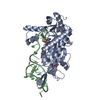
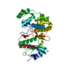
| Title | Structural insights of the Mycoplasma hyorhinis protein Mh-p37: A putative thiamine pyrophosphate transporter | ||||||
 Components Components | High affinity transport system protein p37 | ||||||
 Keywords Keywords | TPP BINDING PROTEIN / Mycoplasma / p37 / TPP / cell membrane / Lipoprotein / membrane / transport / transport protein / Palmitate / extracytoplasmic thiamine binding lipoprotein / Cypl | ||||||
| Function / homology |  Function and homology information Function and homology informationregulation of protein phosphorylation / regulation of cell migration / host cell cytoplasm / plasma membrane Similarity search - Function | ||||||
| Biological species |  Mycoplasma hyorhinis (bacteria) Mycoplasma hyorhinis (bacteria) | ||||||
| Method |  X-RAY DIFFRACTION / SYNCHROTRON / MOLECULAR REPLACEMENT / Resolution: 1.6 Å X-RAY DIFFRACTION / SYNCHROTRON / MOLECULAR REPLACEMENT / Resolution: 1.6 Å | ||||||
 Authors Authors | Sippel, K.H. / Robbins, A.H. / Reutzel, R. / McKenna, R. | ||||||
 Citation Citation | Journal: J.Bacteriol. / Year: 2009 Title: Structural insights into the extracytoplasmic thiamine-binding lipoprotein p37 of Mycoplasma hyorhinis Authors: Sippel, K.H. / Robbins, A.H. / Reutzel, R. / Boehlein, S.K. / Namiki, K. / Goodison, S. / Agbandje-McKenna, M. / Rosser, C.J. / McKenna, R. #1: Journal: Acta Crystallogr.,Sect.D / Year: 2008Title: Structure determination of the cancer-associated mycoplasma hyorhinis protein Mh-p37 Authors: Sippel, K.H. / Robbins, A.H. / Reutzal, R. / Domsic, J. / Boehlein, S.K. / Govindasamy, L. / Agbandje-McKenna, M. / Rosser, C.J. / McKenna, R. | ||||||
| History |
|
- Structure visualization
| Structure viewer | Molecule: MolmilJmol/JSmol |
|---|
- Downloads & links
-Download
| PDBx/mmCIF format | 3eki.cif.gz | 97.6 KB | Display | PDBx/mmCIF format |
|---|---|---|---|---|
| PDB format | pdb3eki.ent.gz | 72.1 KB | Display | PDB format |
| PDBx/mmJSON format | 3eki.json.gz | Tree view | PDBx/mmJSON format | |
| Others |  Other downloads Other downloads |
-Validation report
| Arichive directory | https://data.pdbj.org/pub/pdb/validation_reports/ek/3ekiftp://data.pdbj.org/pub/pdb/validation_reports/ek/3eki | HTTPS FTP |
|---|
-Related structure data
| Related structure data | 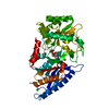 3e78S S: Starting model for refinement |
|---|---|
| Similar structure data |


-Links
 PDBj
PDBj


- Assembly
| Deposited unit | 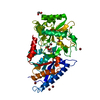
| ||||||||
|---|---|---|---|---|---|---|---|---|---|
| 1 |
| ||||||||
| Unit cell |
| ||||||||
| Details | monomeric |
-Components
-Protein , 1 types, 1 molecules A
| #1: Protein | Mass: 46117.820 Da / Num. of mol.: 1 Source method: isolated from a genetically manipulated source Source: (gene. exp.) Mycoplasma hyorhinis (bacteria) / Gene: p37 / Plasmid: PET31p37 / Production host: |
|---|
-Non-polymers , 5 types, 333 molecules 


| #2: Chemical | ChemComp-TPP /  Mass: 425.314 Da / Num. of mol.: 1 / Source method: obtained synthetically / Formula: C12H19N4O7P2S Mass: 425.314 Da / Num. of mol.: 1 / Source method: obtained synthetically / Formula: C12H19N4O7P2S | ||||
|---|---|---|---|---|---|
| #3: Chemical | ChemComp-GOL /  Mass: 92.094 Da / Num. of mol.: 1 / Source method: obtained synthetically / Formula: C3H8O3 Mass: 92.094 Da / Num. of mol.: 1 / Source method: obtained synthetically / Formula: C3H8O3 | ||||
| #4: Chemical |  Mass: 40.078 Da / Num. of mol.: 2 / Source method: obtained synthetically / Formula: Ca Mass: 40.078 Da / Num. of mol.: 2 / Source method: obtained synthetically / Formula: Ca#5: Chemical | ChemComp-BR /  Mass: 79.904 Da / Num. of mol.: 7 / Source method: obtained synthetically / Formula: Br Mass: 79.904 Da / Num. of mol.: 7 / Source method: obtained synthetically / Formula: Br#6: Water | ChemComp-HOH / | Mass: 18.015 Da / Num. of mol.: 322 / Source method: isolated from a natural source / Formula: H2O |
-Experimental details
-Experiment
| Experiment | Method: X-RAY DIFFRACTION / Number of used crystals: 1 |
|---|
- Sample preparation
| Crystal | Density Matthews: 2.08 Å3/Da / Density % sol: 40.91 % |
|---|---|
| Crystal grow | Temperature: 298 K / Method: batch / pH: 4 Details: 0.1 M Ammonium Bromide, 0.1 M Citric Acid, 40% PEG 4000, under paraffin oil, pH 4.0, Batch, temperature 298K |
-Data collection
| Diffraction | Mean temperature: 100 K |
|---|---|
| Diffraction source | Source: SYNCHROTRON / Site: CHESS  / Beamline: F2 / Wavelength: 0.9169 Å / Beamline: F2 / Wavelength: 0.9169 Å |
| Detector | Type: ADSC QUANTUM 4 / Detector: CCD / Date: Mar 10, 2002 |
| Radiation | Monochromator: Si 111 channel / Protocol: SINGLE WAVELENGTH / Monochromatic (M) / Laue (L): M / Scattering type: x-ray |
| Radiation wavelength | Wavelength: 0.9169 Å / Relative weight: 1 |
| Reflection | Resolution: 1.6→40 Å / Num. all: 49563 / Num. obs: 47500 / % possible obs: 94.6 % / Observed criterion σ(F): 0 / Observed criterion σ(I): 0 / Redundancy: 2.7 % / Rsym value: 0.071 / Net I/σ(I): 0.115 |
| Reflection shell | Resolution: 1.6→1.66 Å / Redundancy: 2.6 % / Mean I/σ(I) obs: 3.1 / Num. unique all: 4759 / Rsym value: 0.288 / % possible all: 95.6 |
- Processing
| Software |
| ||||||||||||||||||||
|---|---|---|---|---|---|---|---|---|---|---|---|---|---|---|---|---|---|---|---|---|---|
| Refinement | Method to determine structure: MOLECULAR REPLACEMENT Starting model: PDB entry 3E78 Resolution: 1.6→40 Å Isotropic thermal model: isotropic except for one calcium ion Cross valid method: THROUGHOUT / σ(F): 0 / σ(I): 0 / Stereochemistry target values: Engh & Huber / Details: Used weighted full matrix least squares procedure.
| ||||||||||||||||||||
| Displacement parameters | Biso mean: 20 Å2 | ||||||||||||||||||||
| Refinement step | Cycle: LAST / Resolution: 1.6→40 Å
| ||||||||||||||||||||
| Refine LS restraints |
|
