 Movie
Movie Controller

[English] 日本語
 Yorodumi
- PDB-3k1x: Acidic Fibroblast Growth Factor (FGF-1) complexed with dobesilate -
+ Open data

- Basic information
| Entry | Database: PDB / ID: 3k1x | ||||||
|---|---|---|---|---|---|---|---|
| Title | Acidic Fibroblast Growth Factor (FGF-1) complexed with dobesilate | ||||||
 Components Components | Heparin-binding growth factor 1 | ||||||
 Keywords Keywords | HORMONE / acidic fibroblast growth factor / inhibitors / ACETYLATION | ||||||
| Function / homology |  Function and homology information Function and homology informationmesonephric epithelium development / branch elongation involved in ureteric bud branching / organ induction / regulation of endothelial tube morphogenesis / FGFR3b ligand binding and activation / regulation of endothelial cell chemotaxis to fibroblast growth factor / Signaling by activated point mutants of FGFR3 / FGFR3c ligand binding and activation / Phospholipase C-mediated cascade; FGFR3 / fibroblast growth factor receptor binding ...mesonephric epithelium development / branch elongation involved in ureteric bud branching / organ induction / regulation of endothelial tube morphogenesis / FGFR3b ligand binding and activation / regulation of endothelial cell chemotaxis to fibroblast growth factor / Signaling by activated point mutants of FGFR3 / FGFR3c ligand binding and activation / Phospholipase C-mediated cascade; FGFR3 / fibroblast growth factor receptor binding / FGFR2b ligand binding and activation / FGFR2c ligand binding and activation / Activated point mutants of FGFR2 / FGFR4 ligand binding and activation / Phospholipase C-mediated cascade; FGFR2 / FGFR1b ligand binding and activation / Phospholipase C-mediated cascade; FGFR4 / Signaling by activated point mutants of FGFR1 / FGFR1c ligand binding and activation / Downstream signaling of activated FGFR1 / Phospholipase C-mediated cascade: FGFR1 / S100 protein binding / activation of protein kinase B activity / Signaling by FGFR2 IIIa TM / PI-3K cascade:FGFR3 / PI-3K cascade:FGFR2 / PI-3K cascade:FGFR4 / positive regulation of sprouting angiogenesis / PI-3K cascade:FGFR1 / positive regulation of MAP kinase activity / fibroblast growth factor receptor signaling pathway / positive regulation of cell division / positive regulation of intracellular signal transduction / PI3K Cascade / anatomical structure morphogenesis / SHC-mediated cascade:FGFR3 / SHC-mediated cascade:FGFR2 / SHC-mediated cascade:FGFR4 / epithelial cell proliferation / SHC-mediated cascade:FGFR1 / FRS-mediated FGFR3 signaling / FRS-mediated FGFR2 signaling / FRS-mediated FGFR4 signaling / FRS-mediated FGFR1 signaling / Signaling by FGFR3 in disease / Signaling by FGFR2 in disease / lung development / neurogenesis / Signaling by FGFR1 in disease / positive regulation of endothelial cell migration / positive regulation of epithelial cell proliferation / regulation of cell migration / growth factor activity / wound healing / Negative regulation of FGFR3 signaling / Negative regulation of FGFR2 signaling / Negative regulation of FGFR4 signaling / Negative regulation of FGFR1 signaling / positive regulation of cholesterol biosynthetic process / integrin binding / Constitutive Signaling by Aberrant PI3K in Cancer / positive regulation of angiogenesis / PIP3 activates AKT signaling / heparin binding / cellular response to heat / PI5P, PP2A and IER3 Regulate PI3K/AKT Signaling / extracellular matrix / RAF/MAP kinase cascade / angiogenesis / cell cortex / positive regulation of MAPK cascade / positive regulation of ERK1 and ERK2 cascade / positive regulation of cell migration / positive regulation of cell population proliferation / signal transduction / positive regulation of transcription by RNA polymerase II / : / extracellular region / nucleoplasm / nucleus / cytosol / cytoplasm Similarity search - Function | ||||||
| Biological species |  Homo sapiens (human) Homo sapiens (human) | ||||||
| Method |  X-RAY DIFFRACTION / SYNCHROTRON / MOLECULAR REPLACEMENT / Resolution: 1.98 Å X-RAY DIFFRACTION / SYNCHROTRON / MOLECULAR REPLACEMENT / Resolution: 1.98 Å | ||||||
 Authors Authors | Romero, A. / Fernandez, I.S. / Gimenez-Gallego, G. | ||||||
 Citation Citation | Journal: J.Biol.Chem. / Year: 2010 Title: Gentisic acid, a compound associated with plant defense and a metabolite of aspirin, heads a new class of in vivo fibroblast growth factor inhibitors. Authors: Fernandez, I.S. / Cuevas, P. / Angulo, J. / Lopez-Navajas, P. / Canales-Mayordomo, A. / Gonzalez-Corrochano, R. / Lozano, R.M. / Valverde, S. / Jimenez-Barbero, J. / Romero, A. / Gimenez-Gallego, G. | ||||||
| History |
|
- Structure visualization
| Structure viewer | Molecule: MolmilJmol/JSmol |
|---|
- Downloads & links
-Download
| PDBx/mmCIF format | 3k1x.cif.gz | 164.7 KB | Display | PDBx/mmCIF format |
|---|---|---|---|---|
| PDB format | pdb3k1x.ent.gz | 133.3 KB | Display | PDB format |
| PDBx/mmJSON format | 3k1x.json.gz | Tree view | PDBx/mmJSON format | |
| Others |  Other downloads Other downloads |
-Validation report
| Arichive directory | https://data.pdbj.org/pub/pdb/validation_reports/k1/3k1xftp://data.pdbj.org/pub/pdb/validation_reports/k1/3k1x | HTTPS FTP |
|---|
-Related structure data
| Related structure data | 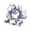 3jutC  1axmS C: citing same article ( S: Starting model for refinement |
|---|---|
| Similar structure data |


-Links
 PDBj
PDBj


- Assembly
| Deposited unit | 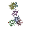
| ||||||||
|---|---|---|---|---|---|---|---|---|---|
| 1 | 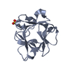
| ||||||||
| 2 | 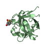
| ||||||||
| 3 | 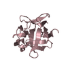
| ||||||||
| 4 | 
| ||||||||
| 5 | 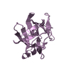
| ||||||||
| 6 | 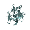
| ||||||||
| Unit cell |
|
-Components
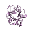
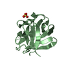
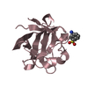
| #1: Protein | Mass: 14752.703 Da / Num. of mol.: 6 / Fragment: Heparin-binding, UNP residues 24-153 Source method: isolated from a genetically manipulated source Source: (gene. exp.) Homo sapiens (human) / Plasmid: pRAT / Production host:  #2: Chemical | 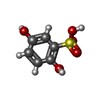  Mass: 190.174 Da / Num. of mol.: 2 / Source method: obtained synthetically / Formula: C6H6O5S Mass: 190.174 Da / Num. of mol.: 2 / Source method: obtained synthetically / Formula: C6H6O5S#3: Water | ChemComp-HOH / |  Mass: 18.015 Da / Num. of mol.: 154 / Source method: isolated from a natural source / Formula: H2O Mass: 18.015 Da / Num. of mol.: 154 / Source method: isolated from a natural source / Formula: H2O |
|---|
-Experimental details
-Experiment
| Experiment | Method: X-RAY DIFFRACTION / Number of used crystals: 1 |
|---|
- Sample preparation
| Crystal | Density Matthews: 2.44 Å3/Da / Density % sol: 49.51 % |
|---|---|
| Crystal grow | Temperature: 295 K / Method: vapor diffusion, sitting drop / pH: 7.8 Details: Crystals of thecomplex between FGF-1 and 2,5-DHPS (2,5-dihydroxyphenylsulfonate) were grown using the sitting drop vapour method at 295 K. Equal volumes of protein and inhibitor solutions, 0. ...Details: Crystals of thecomplex between FGF-1 and 2,5-DHPS (2,5-dihydroxyphenylsulfonate) were grown using the sitting drop vapour method at 295 K. Equal volumes of protein and inhibitor solutions, 0.75 and 1.5mM, respectively were mixed with drops containing 60% sodium/potassium tartrate buffered with 5mM sodium phosphate [pH 7.8]. The drops were equilibrated against 0.2ml of 1.3M Li2SO4 and typical crystals grew within two weeks with approximate dimensions of 0.7 x 0.5 x 0.2 mm. , VAPOR DIFFUSION, SITTING DROP |
-Data collection
| Diffraction | Mean temperature: 100 K |
|---|---|
| Diffraction source | Source: SYNCHROTRON / Site: ESRF  / Beamline: BM16 / Wavelength: 0.979 Å / Beamline: BM16 / Wavelength: 0.979 Å |
| Detector | Type: ADSC QUANTUM 210r / Detector: CCD / Date: Dec 7, 2008 |
| Radiation | Monochromator: Si 111 CHANNEL / Protocol: SINGLE WAVELENGTH / Monochromatic (M) / Laue (L): M / Scattering type: x-ray |
| Radiation wavelength | Wavelength: 0.979 Å / Relative weight: 1 |
| Reflection | Resolution: 1.98→94.07 Å / Num. all: 59910 / Num. obs: 59016 / % possible obs: 98.6 % / Observed criterion σ(F): 0 / Observed criterion σ(I): 0 / Redundancy: 3.7 % / Biso Wilson estimate: 29.604 Å2 / Rmerge(I) obs: 0.05 / Net I/σ(I): 16 |
| Reflection shell | Resolution: 1.98→2.09 Å / Redundancy: 3.3 % / Rmerge(I) obs: 0.342 / Mean I/σ(I) obs: 4.5 / Num. unique all: 8011 / % possible all: 92.8 |
- Processing
| Software |
| ||||||||||||||||||||||||||||||||||||||||||||||||||||||||||||||||||||||||||||||||||||||||||
|---|---|---|---|---|---|---|---|---|---|---|---|---|---|---|---|---|---|---|---|---|---|---|---|---|---|---|---|---|---|---|---|---|---|---|---|---|---|---|---|---|---|---|---|---|---|---|---|---|---|---|---|---|---|---|---|---|---|---|---|---|---|---|---|---|---|---|---|---|---|---|---|---|---|---|---|---|---|---|---|---|---|---|---|---|---|---|---|---|---|---|---|
| Refinement | Method to determine structure: MOLECULAR REPLACEMENT Starting model: PDB ENTRY 1AXM Resolution: 1.98→24.48 Å / Cor.coef. Fo:Fc: 0.946 / Cor.coef. Fo:Fc free: 0.91 / SU B: 5.774 / SU ML: 0.162 / Cross valid method: THROUGHOUT / σ(F): 0 / ESU R: 0.211 / ESU R Free: 0.198 / Stereochemistry target values: MAXIMUM LIKELIHOOD
| ||||||||||||||||||||||||||||||||||||||||||||||||||||||||||||||||||||||||||||||||||||||||||
| Solvent computation | Ion probe radii: 0.8 Å / Shrinkage radii: 0.8 Å / VDW probe radii: 1.2 Å / Solvent model: MASK | ||||||||||||||||||||||||||||||||||||||||||||||||||||||||||||||||||||||||||||||||||||||||||
| Displacement parameters | Biso mean: 36.077 Å2
| ||||||||||||||||||||||||||||||||||||||||||||||||||||||||||||||||||||||||||||||||||||||||||
| Refinement step | Cycle: LAST / Resolution: 1.98→24.48 Å
| ||||||||||||||||||||||||||||||||||||||||||||||||||||||||||||||||||||||||||||||||||||||||||
| Refine LS restraints |
| ||||||||||||||||||||||||||||||||||||||||||||||||||||||||||||||||||||||||||||||||||||||||||
| LS refinement shell | Resolution: 1.981→2.032 Å / Total num. of bins used: 20
|
