 Movie
Movie Controller

[English] 日本語
 Yorodumi
- PDB-3i5u: Crystal structure of an O-methyltransferase (NcsB1) from neocarzi... -
+ Open data

- Basic information
| Entry | Database: PDB / ID: 3i5u | ||||||
|---|---|---|---|---|---|---|---|
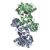
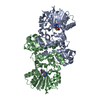
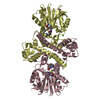
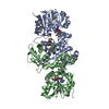
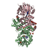
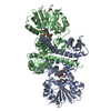
| Title | Crystal structure of an O-methyltransferase (NcsB1) from neocarzinostatin biosynthesis in complex with S-adenosylmethionine (SAM) and 2-hydroxy-5-methyl naphthoic acid (MNA) | ||||||
 Components Components | O-methyltransferase | ||||||
 Keywords Keywords | TRANSFERASE / co-complex / Rossmann-like fold / Methyltransferase | ||||||
| Function / homology |  Function and homology information Function and homology information2,7-dihydroxy-5-methyl-1-naphthoate 7-O-methyltransferase / O-methyltransferase activity / antibiotic biosynthetic process / methylation / protein dimerization activity Similarity search - Function | ||||||
| Biological species |  Streptomyces carzinostaticus subsp. neocarzinostaticus (bacteria) Streptomyces carzinostaticus subsp. neocarzinostaticus (bacteria) | ||||||
| Method |  X-RAY DIFFRACTION / SYNCHROTRON / MOLECULAR REPLACEMENT / Resolution: 2.6 Å X-RAY DIFFRACTION / SYNCHROTRON / MOLECULAR REPLACEMENT / Resolution: 2.6 Å | ||||||
 Authors Authors | Cooke, H.A. / Bruner, S.D. | ||||||
 Citation Citation | Journal: Biochemistry / Year: 2009 Title: Molecular basis of substrate promiscuity for the SAM-dependent O-methyltransferase NcsB1, involved in the biosynthesis of the enediyne antitumor antibiotic neocarzinostatin. Authors: Cooke, H.A. / Guenther, E.L. / Luo, Y. / Shen, B. / Bruner, S.D. | ||||||
| History |
|
- Structure visualization
| Structure viewer | Molecule: MolmilJmol/JSmol |
|---|
- Downloads & links
-Download
| PDBx/mmCIF format | 3i5u.cif.gz | 136.7 KB | Display | PDBx/mmCIF format |
|---|---|---|---|---|
| PDB format | pdb3i5u.ent.gz | 106.7 KB | Display | PDB format |
| PDBx/mmJSON format | 3i5u.json.gz | Tree view | PDBx/mmJSON format | |
| Others |  Other downloads Other downloads |
-Validation report
| Arichive directory | https://data.pdbj.org/pub/pdb/validation_reports/i5/3i5uftp://data.pdbj.org/pub/pdb/validation_reports/i5/3i5u | HTTPS FTP |
|---|
-Related structure data
| Related structure data | 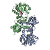 3i53C 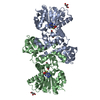 3i58C  3i64C 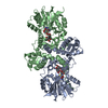 1tw2S C: citing same article ( S: Starting model for refinement |
|---|---|
| Similar structure data |


-Links
 PDBj
PDBj
- Assembly
| Deposited unit | 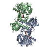
| ||||||||
|---|---|---|---|---|---|---|---|---|---|
| 1 |
| ||||||||
| Unit cell |
|
-Components
| #1: Protein | Mass: 34633.105 Da / Num. of mol.: 2 Source method: isolated from a genetically manipulated source Source: (gene. exp.) Streptomyces carzinostaticus subsp. neocarzinostaticus (bacteria)Gene: ncsb1 / Plasmid: pBS5002 / Production host: References: UniProt: Q84HC8, Transferases; Transferring one-carbon groups; Methyltransferases #2: Chemical |   Mass: 398.437 Da / Num. of mol.: 2 / Source method: obtained synthetically / Formula: C15H22N6O5S Mass: 398.437 Da / Num. of mol.: 2 / Source method: obtained synthetically / Formula: C15H22N6O5S#3: Chemical |   Mass: 202.206 Da / Num. of mol.: 2 / Source method: obtained synthetically / Formula: C12H10O3 Mass: 202.206 Da / Num. of mol.: 2 / Source method: obtained synthetically / Formula: C12H10O3#4: Chemical |   Mass: 92.094 Da / Num. of mol.: 2 / Source method: obtained synthetically / Formula: C3H8O3 Mass: 92.094 Da / Num. of mol.: 2 / Source method: obtained synthetically / Formula: C3H8O3#5: Water | ChemComp-HOH / |  Mass: 18.015 Da / Num. of mol.: 249 / Source method: isolated from a natural source / Formula: H2O Mass: 18.015 Da / Num. of mol.: 249 / Source method: isolated from a natural source / Formula: H2O |
|---|
-Experimental details
-Experiment
| Experiment | Method: X-RAY DIFFRACTION / Number of used crystals: 1 |
|---|
- Sample preparation
| Crystal | Density Matthews: 5.13 Å3/Da / Density % sol: 76.02 % |
|---|---|
| Crystal grow | Temperature: 293 K / Method: vapor diffusion, hanging drop / pH: 3.8 Details: 3.6 M Sodium formate, 10% Glycerol, 2 mM SAM, 2 mM MNA, pH 3.8, VAPOR DIFFUSION, HANGING DROP, temperature 293K |
-Data collection
| Diffraction | Mean temperature: 100 K |
|---|---|
| Diffraction source | Source: SYNCHROTRON / Site: NSLS  / Beamline: X25 / Wavelength: 1 Å / Beamline: X25 / Wavelength: 1 Å |
| Detector | Type: ADSC QUANTUM 315 / Detector: CCD / Date: Nov 14, 2008 |
| Radiation | Monochromator: Si(111) crystal / Protocol: SINGLE WAVELENGTH / Monochromatic (M) / Laue (L): M / Scattering type: x-ray |
| Radiation wavelength | Wavelength: 1 Å / Relative weight: 1 |
| Reflection | Resolution: 2.6→50 Å / Num. obs: 42568 / % possible obs: 99.8 % / Redundancy: 22.7 % / Biso Wilson estimate: 49.7 Å2 / Rmerge(I) obs: 0.077 / Net I/σ(I): 49.6 |
| Reflection shell | Resolution: 2.6→2.69 Å / Redundancy: 22.9 % / Rmerge(I) obs: 0.441 / Mean I/σ(I) obs: 6.4 / Num. unique all: 4243 / % possible all: 100 |
- Processing
| Software |
| |||||||||||||||||||||||||
|---|---|---|---|---|---|---|---|---|---|---|---|---|---|---|---|---|---|---|---|---|---|---|---|---|---|---|
| Refinement | Method to determine structure: MOLECULAR REPLACEMENT Starting model: PDB entry 1TW2 Resolution: 2.6→30.13 Å / Cross valid method: FREE R
| |||||||||||||||||||||||||
| Displacement parameters | Biso mean: 55.2 Å2
| |||||||||||||||||||||||||
| Refine analyze |
| |||||||||||||||||||||||||
| Refinement step | Cycle: LAST / Resolution: 2.6→30.13 Å
| |||||||||||||||||||||||||
| Refine LS restraints |
| |||||||||||||||||||||||||
| LS refinement shell | Resolution: 2.6→2.76 Å / Rfactor Rfree error: 0.02 / Total num. of bins used: 10
|
