 Movie
Movie Controller

[English] 日本語
 Yorodumi
- PDB-3ert: HUMAN ESTROGEN RECEPTOR ALPHA LIGAND-BINDING DOMAIN IN COMPLEX WI... -
+ Open data

- Basic information
| Entry | Database: PDB / ID: 3ert | |||||||||
|---|---|---|---|---|---|---|---|---|---|---|
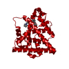
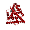
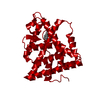
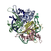
| Title | HUMAN ESTROGEN RECEPTOR ALPHA LIGAND-BINDING DOMAIN IN COMPLEX WITH 4-HYDROXYTAMOXIFEN | |||||||||
 Components Components | PROTEIN (ESTROGEN RECEPTOR ALPHA) | |||||||||
 Keywords Keywords | NUCLEAR RECEPTOR / TRANSCRIPTION FACTOR / ESTROGEN / ANTAGONIST | |||||||||
| Function / homology |  Function and homology information Function and homology informationregulation of epithelial cell apoptotic process / steroid hormone receptor signaling pathway / antral ovarian follicle growth / regulation of branching involved in prostate gland morphogenesis / RUNX1 regulates transcription of genes involved in WNT signaling / RUNX1 regulates estrogen receptor mediated transcription / nuclear estrogen receptor activity / regulation of toll-like receptor signaling pathway / prostate epithelial cord elongation / epithelial cell proliferation involved in mammary gland duct elongation ...regulation of epithelial cell apoptotic process / steroid hormone receptor signaling pathway / antral ovarian follicle growth / regulation of branching involved in prostate gland morphogenesis / RUNX1 regulates transcription of genes involved in WNT signaling / RUNX1 regulates estrogen receptor mediated transcription / nuclear estrogen receptor activity / regulation of toll-like receptor signaling pathway / prostate epithelial cord elongation / epithelial cell proliferation involved in mammary gland duct elongation / prostate epithelial cord arborization involved in prostate glandular acinus morphogenesis / epithelial cell development / mammary gland branching involved in pregnancy / uterus development / vagina development / TFIIB-class transcription factor binding / androgen metabolic process / negative regulation of smooth muscle cell apoptotic process / nuclear receptor-mediated steroid hormone signaling pathway / mammary gland alveolus development / : / cellular response to estrogen stimulus / estrogen response element binding / : / Mitochondrial unfolded protein response (UPRmt) / Nuclear signaling by ERBB4 / RNA polymerase II preinitiation complex assembly / positive regulation of nitric-oxide synthase activity / estrogen receptor signaling pathway / steroid binding / TFAP2 (AP-2) family regulates transcription of growth factors and their receptors / protein localization to chromatin / 14-3-3 protein binding / ESR-mediated signaling / negative regulation of miRNA transcription / TBP-class protein binding / nitric-oxide synthase regulator activity / nuclear estrogen receptor binding / transcription corepressor binding / transcription coregulator binding / stem cell differentiation / negative regulation of canonical NF-kappaB signal transduction / SUMOylation of intracellular receptors / cellular response to estradiol stimulus / euchromatin / Nuclear Receptor transcription pathway / response to estrogen / beta-catenin binding / male gonad development / nuclear receptor activity / positive regulation of fibroblast proliferation / transcription coactivator binding / Constitutive Signaling by Aberrant PI3K in Cancer / positive regulation of nitric oxide biosynthetic process / sequence-specific double-stranded DNA binding / Regulation of RUNX2 expression and activity / Ovarian tumor domain proteases / response to estradiol / PIP3 activates AKT signaling / ATPase binding / PI5P, PP2A and IER3 Regulate PI3K/AKT Signaling / positive regulation of cytosolic calcium ion concentration / regulation of inflammatory response / fibroblast proliferation / DNA-binding transcription activator activity, RNA polymerase II-specific / transcription regulator complex / Estrogen-dependent gene expression / phospholipase C-activating G protein-coupled receptor signaling pathway / DNA-binding transcription factor activity, RNA polymerase II-specific / calmodulin binding / Extra-nuclear estrogen signaling / RNA polymerase II cis-regulatory region sequence-specific DNA binding / chromatin remodeling / DNA-binding transcription factor activity / negative regulation of gene expression / chromatin binding / regulation of transcription by RNA polymerase II / regulation of DNA-templated transcription / protein kinase binding / positive regulation of DNA-templated transcription / chromatin / enzyme binding / negative regulation of transcription by RNA polymerase II / Golgi apparatus / signal transduction / positive regulation of transcription by RNA polymerase II / protein-containing complex / zinc ion binding / nucleoplasm / membrane / identical protein binding / nucleus / plasma membrane / cytoplasm / cytosol Similarity search - Function | |||||||||
| Biological species |  Homo sapiens (human) Homo sapiens (human) | |||||||||
| Method |  X-RAY DIFFRACTION / SYNCHROTRON / MOLECULAR REPLACEMENT / Resolution: 1.9 Å X-RAY DIFFRACTION / SYNCHROTRON / MOLECULAR REPLACEMENT / Resolution: 1.9 Å | |||||||||
 Authors Authors | Shiau, A.K. / Barstad, D. / Loria, P.M. / Cheng, L. / Kushner, P.J. / Agard, D.A. / Greene, G.L. | |||||||||
 Citation Citation | Journal: Cell(Cambridge,Mass.) / Year: 1998 Title: The structural basis of estrogen receptor/coactivator recognition and the antagonism of this interaction by tamoxifen. Authors: Shiau, A.K. / Barstad, D. / Loria, P.M. / Cheng, L. / Kushner, P.J. / Agard, D.A. / Greene, G.L. | |||||||||
| History |
|
- Structure visualization
| Structure viewer | Molecule: MolmilJmol/JSmol |
|---|
- Downloads & links
-Download
| PDBx/mmCIF format | 3ert.cif.gz | 64.9 KB | Display | PDBx/mmCIF format |
|---|---|---|---|---|
| PDB format | pdb3ert.ent.gz | 46.9 KB | Display | PDB format |
| PDBx/mmJSON format | 3ert.json.gz | Tree view | PDBx/mmJSON format | |
| Others |  Other downloads Other downloads |
-Validation report
| Arichive directory | https://data.pdbj.org/pub/pdb/validation_reports/er/3ertftp://data.pdbj.org/pub/pdb/validation_reports/er/3ert | HTTPS FTP |
|---|
-Related structure data
| Related structure data | 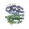 3erdC 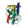 2lbdS S: Starting model for refinement C: citing same article ( |
|---|---|
| Similar structure data |


-Links
 PDBj
PDBj


- Assembly
| Deposited unit | 
| |||||||||
|---|---|---|---|---|---|---|---|---|---|---|
| 1 | 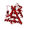
| |||||||||
| Unit cell |
| |||||||||
| Components on special symmetry positions |
|
-Components
| #1: Protein | Mass: 29850.217 Da / Num. of mol.: 1 / Fragment: LIGAND-BINDING DOMAIN Source method: isolated from a genetically manipulated source Source: (gene. exp.) Homo sapiens (human) / Gene: ESTROGEN RECEPTOR ALPHA / Plasmid: PET23D / Cellular location (production host): CYTOPLASM / Production host:  |
|---|---|
| #2: Chemical | ChemComp-OHT /   Mass: 387.514 Da / Num. of mol.: 1 / Source method: obtained synthetically / Formula: C26H29NO2 Mass: 387.514 Da / Num. of mol.: 1 / Source method: obtained synthetically / Formula: C26H29NO2 |
| #3: Water | ChemComp-HOH /  Mass: 18.015 Da / Num. of mol.: 79 / Source method: isolated from a natural source / Formula: H2O Mass: 18.015 Da / Num. of mol.: 79 / Source method: isolated from a natural source / Formula: H2O |
-Experimental details
-Experiment
| Experiment | Method: X-RAY DIFFRACTION / Number of used crystals: 1 |
|---|
- Sample preparation
| Crystal | Density Matthews: 2.4 Å3/Da / Density % sol: 48 % | |||||||||||||||||||||||||
|---|---|---|---|---|---|---|---|---|---|---|---|---|---|---|---|---|---|---|---|---|---|---|---|---|---|---|
| Crystal grow | pH: 7 Details: WELL: 25-27%(W/V) PEG 4000, 0.180 M SODIUM ACETATE, 0.90 M TRIS PH 8.75-9.0 PROTEIN: 4.3 G/L TEMPERATURE: 19-21 DEGREES C, pH 7.0 | |||||||||||||||||||||||||
| Crystal grow | *PLUS Temperature: 19-21 ℃ / Method: vapor diffusion, hanging drop | |||||||||||||||||||||||||
| Components of the solutions | *PLUS
|
-Data collection
| Diffraction | Mean temperature: 100 K |
|---|---|
| Diffraction source | Source: SYNCHROTRON / Site: SSRL  / Beamline: BL9-1 / Wavelength: 0.98 / Beamline: BL9-1 / Wavelength: 0.98 |
| Detector | Type: MARRESEARCH / Detector: IMAGE PLATE / Date: Mar 1, 1998 |
| Radiation | Protocol: SINGLE WAVELENGTH / Monochromatic (M) / Laue (L): M / Scattering type: x-ray |
| Radiation wavelength | Wavelength: 0.98 Å / Relative weight: 1 |
| Reflection | Resolution: 1.9→41 Å / Num. obs: 23064 / % possible obs: 99.1 % / Observed criterion σ(I): -3 / Redundancy: 11.7 % / Biso Wilson estimate: 33.7 Å2 / Rsym value: 0.07 / Net I/σ(I): 16.1 |
| Reflection shell | Resolution: 1.9→1.93 Å / Mean I/σ(I) obs: 2.1 / Rsym value: 0.646 / % possible all: 97.1 |
| Reflection | *PLUS Num. measured all: 269253 / Rmerge(I) obs: 0.07 |
| Reflection shell | *PLUS % possible obs: 97.1 % / Rmerge(I) obs: 0.646 |
- Processing
| Software |
| ||||||||||||||||||||||||||||||||||||||||||||||||||||||||||||
|---|---|---|---|---|---|---|---|---|---|---|---|---|---|---|---|---|---|---|---|---|---|---|---|---|---|---|---|---|---|---|---|---|---|---|---|---|---|---|---|---|---|---|---|---|---|---|---|---|---|---|---|---|---|---|---|---|---|---|---|---|---|
| Refinement | Method to determine structure: MOLECULAR REPLACEMENT Starting model: 2LBD Resolution: 1.9→41 Å / Data cutoff high absF: 10000000 / Data cutoff low absF: 0.001 / Cross valid method: THROUGHOUT / σ(F): 0
| ||||||||||||||||||||||||||||||||||||||||||||||||||||||||||||
| Displacement parameters | Biso mean: 40.4 Å2
| ||||||||||||||||||||||||||||||||||||||||||||||||||||||||||||
| Refinement step | Cycle: LAST / Resolution: 1.9→41 Å
| ||||||||||||||||||||||||||||||||||||||||||||||||||||||||||||
| Refine LS restraints |
| ||||||||||||||||||||||||||||||||||||||||||||||||||||||||||||
| LS refinement shell | Resolution: 1.9→1.99 Å / Total num. of bins used: 8
| ||||||||||||||||||||||||||||||||||||||||||||||||||||||||||||
| Xplor file |
| ||||||||||||||||||||||||||||||||||||||||||||||||||||||||||||
| Software | *PLUS Name: X-PLOR / Version: 3.854 / Classification: refinement | ||||||||||||||||||||||||||||||||||||||||||||||||||||||||||||
| Refinement | *PLUS Rfactor obs: 0.229 | ||||||||||||||||||||||||||||||||||||||||||||||||||||||||||||
| Solvent computation | *PLUS | ||||||||||||||||||||||||||||||||||||||||||||||||||||||||||||
| Displacement parameters | *PLUS | ||||||||||||||||||||||||||||||||||||||||||||||||||||||||||||
| Refine LS restraints | *PLUS
| ||||||||||||||||||||||||||||||||||||||||||||||||||||||||||||
| LS refinement shell | *PLUS Rfactor obs: 0.365 |
