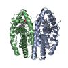
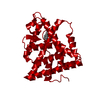
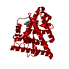
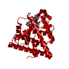
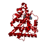
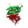
[English] 日本語
Yorodumi
- PDB-2ouz: Crystal Structure of Estrogen Receptor alpha-lasofoxifene complex -

+
Open data


ID or keywords:

Loading...

-
Basic information

Entry
Database: PDB / ID: 2ouz
TitleCrystal Structure of Estrogen Receptor alpha-lasofoxifene complex
ComponentsEstrogen receptor
KeywordsHORMONE/GROWTH FACTOR / Nuclear receptor / SERM / estrogen / HORMONE-GROWTH FACTOR COMPLEX
Function / homology
Function and homology information


regulation of epithelial cell apoptotic process / antral ovarian follicle growth / G protein-coupled estrogen receptor activity / regulation of branching involved in prostate gland morphogenesis / RUNX1 regulates transcription of genes involved in WNT signaling / RUNX1 regulates estrogen receptor mediated transcription / regulation of toll-like receptor signaling pathway / prostate epithelial cord arborization involved in prostate glandular acinus morphogenesis / nuclear estrogen receptor activity / epithelial cell proliferation involved in mammary gland duct elongation ...regulation of epithelial cell apoptotic process / antral ovarian follicle growth / G protein-coupled estrogen receptor activity / regulation of branching involved in prostate gland morphogenesis / RUNX1 regulates transcription of genes involved in WNT signaling / RUNX1 regulates estrogen receptor mediated transcription / regulation of toll-like receptor signaling pathway / prostate epithelial cord arborization involved in prostate glandular acinus morphogenesis / nuclear estrogen receptor activity / epithelial cell proliferation involved in mammary gland duct elongation / epithelial cell development / prostate epithelial cord elongation / negative regulation of smooth muscle cell apoptotic process / mammary gland branching involved in pregnancy / uterus development / vagina development / TFIIB-class transcription factor binding / androgen metabolic process / steroid hormone receptor signaling pathway / mammary gland alveolus development / estrogen receptor signaling pathway / cellular response to estrogen stimulus / estrogen response element binding / : / nuclear receptor-mediated steroid hormone signaling pathway / negative regulation of canonical NF-kappaB signal transduction / Nuclear signaling by ERBB4 / RNA polymerase II preinitiation complex assembly / protein localization to chromatin / 14-3-3 protein binding / TBP-class protein binding / nitric-oxide synthase regulator activity / steroid binding / TFAP2 (AP-2) family regulates transcription of growth factors and their receptors / ESR-mediated signaling / transcription corepressor binding / negative regulation of miRNA transcription / positive regulation of nitric-oxide synthase activity / cellular response to estradiol stimulus / transcription coregulator binding / stem cell differentiation / nuclear estrogen receptor binding / SUMOylation of intracellular receptors / euchromatin / negative regulation of DNA-binding transcription factor activity / beta-catenin binding / transcription coactivator binding / positive regulation of DNA-binding transcription factor activity / Nuclear Receptor transcription pathway / response to estrogen / Regulation of RUNX2 expression and activity / Constitutive Signaling by Aberrant PI3K in Cancer / nuclear receptor activity / male gonad development / positive regulation of nitric oxide biosynthetic process / sequence-specific double-stranded DNA binding / positive regulation of fibroblast proliferation / Ovarian tumor domain proteases / PIP3 activates AKT signaling / response to estradiol / phospholipase C-activating G protein-coupled receptor signaling pathway / positive regulation of cytosolic calcium ion concentration / ATPase binding / regulation of inflammatory response / fibroblast proliferation / PI5P, PP2A and IER3 Regulate PI3K/AKT Signaling / DNA-binding transcription activator activity, RNA polymerase II-specific / Estrogen-dependent gene expression / transcription regulator complex / Extra-nuclear estrogen signaling / calmodulin binding / DNA-binding transcription factor activity, RNA polymerase II-specific / chromatin remodeling / RNA polymerase II cis-regulatory region sequence-specific DNA binding / DNA-binding transcription factor activity / negative regulation of gene expression / chromatin binding / regulation of DNA-templated transcription / chromatin / regulation of transcription by RNA polymerase II / protein kinase binding / positive regulation of DNA-templated transcription / Golgi apparatus / enzyme binding / negative regulation of transcription by RNA polymerase II / signal transduction / positive regulation of transcription by RNA polymerase II / protein-containing complex / zinc ion binding / nucleoplasm / identical protein binding / membrane / nucleus / plasma membrane / cytoplasm / cytosol
Similarity search - Function
Estrogen receptor / Oestrogen-type nuclear receptor final C-terminal domain / : / Oestrogen receptor / Oestrogen-type nuclear receptor final C-terminal / Estrogen receptor/oestrogen-related receptor / Retinoid X Receptor / Retinoid X Receptor / Nuclear hormone receptor / Nuclear hormones receptors DNA-binding region signature. ...Estrogen receptor / Oestrogen-type nuclear receptor final C-terminal domain / : / Oestrogen receptor / Oestrogen-type nuclear receptor final C-terminal / Estrogen receptor/oestrogen-related receptor / Retinoid X Receptor / Retinoid X Receptor / Nuclear hormone receptor / Nuclear hormones receptors DNA-binding region signature. / Zinc finger, nuclear hormone receptor-type / Zinc finger, C4 type (two domains) / Nuclear hormone receptors DNA-binding domain profile. / c4 zinc finger in nuclear hormone receptors / Nuclear hormone receptor, ligand-binding domain / Nuclear hormone receptor-like domain superfamily / Ligand-binding domain of nuclear hormone receptor / Nuclear receptor (NR) ligand-binding (LBD) domain profile. / Ligand binding domain of hormone receptors / Zinc finger, NHR/GATA-type / Orthogonal Bundle / Mainly Alpha
Similarity search - Domain/homology
Chem-C3D / Estrogen receptor
Similarity search - Component
Biological speciesHomo sapiens (human)
MethodX-RAY DIFFRACTION / MOLECULAR REPLACEMENT / Resolution: 2 Å
AuthorsVajdos, F.F. / Pandit, J.
CitationJournal: Protein Sci. / Year: 2007
Title: The 2.0 A crystal structure of the ER{alpha} ligand-binding domain complexed with lasofoxifene
Authors: Vajdos, F.F. / Hoth, L.R. / Geoghegan, K.F. / Simons, S.P. / LeMotte, P.K. / Danley, D.E. / Ammirati, M.J. / Pandit, J.
History
DepositionFeb 12, 2007Deposition site: RCSB / Processing site: RCSB
Revision 1.0May 8, 2007Provider: repository / Type: Initial release
Revision 1.1May 1, 2008Group: Version format compliance
Revision 1.2Jul 13, 2011Group: Advisory / Version format compliance
Revision 1.3Apr 4, 2018Group: Data collection / Category: diffrn_source / Item: _diffrn_source.type
Revision 1.4Jan 15, 2020Group: Structure summary / Category: chem_comp / Item: _chem_comp.pdbx_synonyms
Revision 1.5Aug 30, 2023Group: Data collection / Database references ...Data collection / Database references / Derived calculations / Refinement description
Category: chem_comp_atom / chem_comp_bond ...chem_comp_atom / chem_comp_bond / database_2 / pdbx_initial_refinement_model / struct_site
Item: _database_2.pdbx_DOI / _database_2.pdbx_database_accession ..._database_2.pdbx_DOI / _database_2.pdbx_database_accession / _struct_site.pdbx_auth_asym_id / _struct_site.pdbx_auth_comp_id / _struct_site.pdbx_auth_seq_id

-
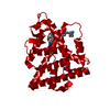
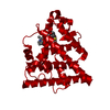
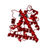
Structure visualization

Structure viewerMolecule:
MolmilJmol/JSmol

Downloads & links

-
Assembly

Deposited unit
A: Estrogen receptor
hetero molecules


Theoretical massNumber of molelcules
Total (without water)29,3032
Polymers28,8891
Non-polymers4141
Water2,900161
1
A: Estrogen receptor
hetero molecules

A: Estrogen receptor
hetero molecules


Theoretical massNumber of molelcules
Total (without water)58,6054
Polymers57,7782
Non-polymers8272
Water362
TypeNameSymmetry operationNumber
identity operation1_555x,y,z1
crystal symmetry operation10_665-y+1,-x+1,-z+1/61
Unit cell
Length a, b, c (Å)58.306, 58.306, 275.033
Angle α, β, γ (deg.)90.000, 90.000, 120.000
Int Tables number179
Space group name H-MP6522

-
Components

#1: Protein Estrogen receptor / ER / Estradiol receptor / ER-alpha


Mass: 28889.021 Da / Num. of mol.: 1 / Fragment: Ligand Binding Domain, residues 306-551
Source method: isolated from a genetically manipulated source
Source: (gene. exp.) Homo sapiens (human) / Gene: ESR1, ESR, NR3A1 / Production host: Escherichia coli (E. coli) / References: UniProt: P03372
#2: Chemical ChemComp-C3D / (5R,6S)-6-PHENYL-5-[4-(2-PYRROLIDIN-1-YLETHOXY)PHENYL]-5,6,7,8-TETRAHYDRONAPHTHALEN-2-OL / Lasofoxifene


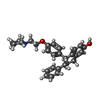
Mass: 413.551 Da / Num. of mol.: 1 / Source method: obtained synthetically / Formula: C28H31NO2
#3: Water ChemComp-HOH / water


Mass: 18.015 Da / Num. of mol.: 161 / Source method: isolated from a natural source / Formula: H2O

-
Experimental details

-
Experiment

ExperimentMethod: X-RAY DIFFRACTION / Number of used crystals: 1

-
Sample preparation

CrystalDensity Matthews: 2.4 Å3/Da / Density % sol: 48.68 %
Crystal growTemperature: 295 K / Method: vapor diffusion, hanging drop / pH: 6.7
Details: 0.1 M NaHEPES, 0.5 M NaCl, 6% ethylene glycol, 10-12%PEG 8000, 5mM DTT, pH 6.7, VAPOR DIFFUSION, HANGING DROP, temperature 295K

-
Data collection

DiffractionMean temperature: 100 K
Diffraction sourceSource: ROTATING ANODE / Type: RIGAKU RU200 / Wavelength: 1.5418 Å
DetectorType: RIGAKU RAXIS IIC / Detector: IMAGE PLATE / Date: May 3, 1999 / Details: osmic
RadiationMonochromator: Multilayer optics / Protocol: SINGLE WAVELENGTH / Monochromatic (M) / Laue (L): M / Scattering type: x-ray
Radiation wavelengthWavelength: 1.5418 Å / Relative weight: 1
ReflectionResolution: 2→50.64 Å / Num. all: 19969 / Num. obs: 18869 / % possible obs: 94.5 % / Observed criterion σ(F): 4 / Observed criterion σ(I): 2 / Redundancy: 3.25 % / Biso Wilson estimate: 41 Å2 / Rmerge(I) obs: 0.073 / Net I/σ(I): 15
Reflection shellResolution: 2→2.07 Å / Rmerge(I) obs: 0.651 / Mean I/σ(I) obs: 1.61 / % possible all: 87.5

-
Processing

Software
NameVersionClassificationNB
REFMAC5.2.0005refinement
PDB_EXTRACT2data extraction
DENZOdata reduction
SCALEPACKdata scaling
AMoREphasing
RefinementMethod to determine structure: MOLECULAR REPLACEMENT
Starting model: PDB entry 3ERT
Resolution: 2→50.64 Å / Cor.coef. Fo:Fc: 0.946 / Cor.coef. Fo:Fc free: 0.901 / SU B: 9.77 / SU ML: 0.14 / TLS residual ADP flag: LIKELY RESIDUAL / Isotropic thermal model: Isotropic restrained / Cross valid method: THROUGHOUT / σ(F): 0 / ESU R: 0.197 / ESU R Free: 0.193 / Stereochemistry target values: MAXIMUM LIKELIHOOD / Details: HYDROGENS HAVE BEEN ADDED IN THE RIDING POSITIONS
RfactorNum. reflection% reflectionSelection details
Rfree0.269 976 5.2 %RANDOM
Rwork0.199 ---
obs0.202 18800 94.64 %-
Solvent computationIon probe radii: 0.8 Å / Shrinkage radii: 0.8 Å / VDW probe radii: 1.4 Å / Solvent model: MASK
Displacement parametersBiso mean: 39.196 Å2
Baniso -1Baniso -2Baniso -3
1-0.74 Å20.37 Å20 Å2
2--0.74 Å20 Å2
3----1.12 Å2
Refine analyzeLuzzati coordinate error obs: 0.308 Å
Refinement stepCycle: LAST / Resolution: 2→50.64 Å
ProteinNucleic acidLigandSolventTotal
Num. atoms1939 0 31 161 2131
Refine LS restraints
Refine-IDTypeDev idealDev ideal targetNumber
X-RAY DIFFRACTIONr_bond_refined_d0.020.0212013
X-RAY DIFFRACTIONr_bond_other_d0.0010.021898
X-RAY DIFFRACTIONr_angle_refined_deg1.7171.9972727
X-RAY DIFFRACTIONr_angle_other_deg0.96134396
X-RAY DIFFRACTIONr_dihedral_angle_1_deg6.0015245
X-RAY DIFFRACTIONr_dihedral_angle_2_deg40.15424.14682
X-RAY DIFFRACTIONr_dihedral_angle_3_deg16.9215364
X-RAY DIFFRACTIONr_dihedral_angle_4_deg18.2481510
X-RAY DIFFRACTIONr_chiral_restr0.1120.2318
X-RAY DIFFRACTIONr_gen_planes_refined0.0070.022172
X-RAY DIFFRACTIONr_gen_planes_other0.0010.02382
X-RAY DIFFRACTIONr_nbd_refined0.2440.2534
X-RAY DIFFRACTIONr_nbd_other0.1850.21967
X-RAY DIFFRACTIONr_nbtor_refined0.1810.2982
X-RAY DIFFRACTIONr_nbtor_other0.0910.21194
X-RAY DIFFRACTIONr_xyhbond_nbd_refined0.2410.2108
X-RAY DIFFRACTIONr_symmetry_vdw_refined0.350.230
X-RAY DIFFRACTIONr_symmetry_vdw_other0.2460.285
X-RAY DIFFRACTIONr_symmetry_hbond_refined0.5490.219
X-RAY DIFFRACTIONr_mcbond_it1.1281.51273
X-RAY DIFFRACTIONr_mcbond_other0.2741.5496
X-RAY DIFFRACTIONr_mcangle_it1.84121971
X-RAY DIFFRACTIONr_scbond_it2.5243847
X-RAY DIFFRACTIONr_scangle_it3.5284.5756
LS refinement shellResolution: 2→2.052 Å / Total num. of bins used: 20
RfactorNum. reflection% reflection
Rfree0.296 71 -
Rwork0.271 1138 -
obs-1209 85.93 %
Refinement TLS params.

Method: refined / Refine-ID: X-RAY DIFFRACTION

IDL112)L122)L132)L222)L232)L332)S11 (Å °)S12 (Å °)S13 (Å °)S21 (Å °)S22 (Å °)S23 (Å °)S31 (Å °)S32 (Å °)S33 (Å °)T112)T122)T132)T222)T232)T332)Origin x (Å)Origin y (Å)Origin z (Å)
11.5117-0.4853-0.17633.1097-0.15482.8193-0.0066-0.0508-0.06550.0392-0.04520.06640.2063-0.11980.0518-0.185-0.09430.0077-0.2176-0.0131-0.198422.02066.479223.1147
220.87230.6013.595911.84584.3256.93620.1048-0.235-0.5558-0.6407-0.1708-0.45810.52490.46550.0660.12260.0161-0.0012-0.1033-0.0085-0.190828.9216-2.51148.881
Refinement TLS group

Refine-ID: X-RAY DIFFRACTION / Selection: ALL / Auth asym-ID: A

IDRefine TLS-IDLabel asym-IDAuth seq-IDLabel seq-ID
11A306 - 5286 - 228
21B9991
32A536 - 544236 - 244

+
About Yorodumi

-
News

-
Feb 9, 2022. New format data for meta-information of EMDB entries

New format data for meta-information of EMDB entries

  • Version 3 of the EMDB header file is now the official format.
  • The previous official version 1.9 will be removed from the archive.

Related info.:EMDB header

External links:wwPDB to switch to version 3 of the EMDB data model

-
Aug 12, 2020. Covid-19 info

Covid-19 info

URL: https://pdbjlvh1.pdbj.org/emnavi/covid19.php

New page: Covid-19 featured information page in EM Navigator.

Related info.:Covid-19 info / Mar 5, 2020. Novel coronavirus structure data

+
Mar 5, 2020. Novel coronavirus structure data

Novel coronavirus structure data

Related info.:Yorodumi Speices / Aug 12, 2020. Covid-19 info

External links:COVID-19 featured content - PDBj / Molecule of the Month (242):Coronavirus Proteases

+
Jan 31, 2019. EMDB accession codes are about to change! (news from PDBe EMDB page)

EMDB accession codes are about to change! (news from PDBe EMDB page)

  • The allocation of 4 digits for EMDB accession codes will soon come to an end. Whilst these codes will remain in use, new EMDB accession codes will include an additional digit and will expand incrementally as the available range of codes is exhausted. The current 4-digit format prefixed with “EMD-” (i.e. EMD-XXXX) will advance to a 5-digit format (i.e. EMD-XXXXX), and so on. It is currently estimated that the 4-digit codes will be depleted around Spring 2019, at which point the 5-digit format will come into force.
  • The EM Navigator/Yorodumi systems omit the EMD- prefix.

Related info.:Q: What is EMD? / ID/Accession-code notation in Yorodumi/EM Navigator

External links:EMDB Accession Codes are Changing Soon! / Contact to PDBj

+
Jul 12, 2017. Major update of PDB

Major update of PDB

  • wwPDB released updated PDB data conforming to the new PDBx/mmCIF dictionary.
  • This is a major update changing the version number from 4 to 5, and with Remediation, in which all the entries are updated.
  • In this update, many items about electron microscopy experimental information are reorganized (e.g. em_software).
  • Now, EM Navigator and Yorodumi are based on the updated data.

External links:wwPDB Remediation / Enriched Model Files Conforming to OneDep Data Standards Now Available in the PDB FTP Archive

-
Yorodumi

Thousand views of thousand structures

  • Yorodumi is a browser for structure data from EMDB, PDB, SASBDB, etc.
  • This page is also the successor to EM Navigator detail page, and also detail information page/front-end page for Omokage search.
  • The word "yorodu" (or yorozu) is an old Japanese word meaning "ten thousand". "mi" (miru) is to see.

Related info.:EMDB / PDB / SASBDB / Comparison of 3 databanks / Yorodumi Search / Aug 31, 2016. New EM Navigator & Yorodumi / Yorodumi Papers / Jmol/JSmol / Function and homology information / Changes in new EM Navigator and Yorodumi

Read more