 Movie
Movie Controller

[English] 日本語
 Yorodumi
- PDB-3eq8: Prolyl oligopeptidase complexed with R-Pro-(decarboxy-Pro)-Type i... -
+ Open data

- Basic information
| Entry | Database: PDB / ID: 3eq8 | ||||||
|---|---|---|---|---|---|---|---|
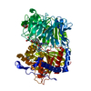
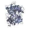
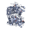
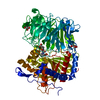
| Title | Prolyl oligopeptidase complexed with R-Pro-(decarboxy-Pro)-Type inhibitors | ||||||
 Components Components | Prolyl endopeptidase | ||||||
 Keywords Keywords | Hydrolase/Hydrolase inhibitor / Hydrolase / Protease / Serine protease / Hydrolase-Hydrolase inhibitor complex | ||||||
| Function / homology |  Function and homology information Function and homology informationprolyl oligopeptidase / serine-type endopeptidase activity / proteolysis / cytoplasm Similarity search - Function | ||||||
| Biological species |  | ||||||
| Method |  X-RAY DIFFRACTION / FOURIER SYNTHESIS / Resolution: 2.73 Å X-RAY DIFFRACTION / FOURIER SYNTHESIS / Resolution: 2.73 Å | ||||||
 Authors Authors | Kanai, K. / Aranyi, P. / Bocskei, Z. / Ferenczy, G. / Harmat, V. / Simon, K. / Naray-Szabo, G. / Hermecz, I. | ||||||
 Citation Citation | Journal: J.Med.Chem. / Year: 2008 Title: Prolyl oligopeptidase inhibition by N-acyl-pro-pyrrolidine-type molecules Authors: Kanai, K. / Aranyi, P. / Bocskei, Z. / Ferenczy, G. / Harmat, V. / Simon, K. / Batori, S. / Naray-Szabo, G. / Hermecz, I. | ||||||
| History |
|
- Structure visualization
| Structure viewer | Molecule: MolmilJmol/JSmol |
|---|
- Downloads & links
-Download
| PDBx/mmCIF format | 3eq8.cif.gz | 141.7 KB | Display | PDBx/mmCIF format |
|---|---|---|---|---|
| PDB format | pdb3eq8.ent.gz | 111.3 KB | Display | PDB format |
| PDBx/mmJSON format | 3eq8.json.gz | Tree view | PDBx/mmJSON format | |
| Others |  Other downloads Other downloads |
-Validation report
| Summary document | 3eq8_validation.pdf.gz | 706.4 KB | Display | wwPDB validaton report |
|---|---|---|---|---|
| Full document | 3eq8_full_validation.pdf.gz | 729.4 KB | Display | |
| Data in XML | 3eq8_validation.xml.gz | 29.3 KB | Display | |
| Data in CIF | 3eq8_validation.cif.gz | 40 KB | Display | |
| Arichive directory | https://data.pdbj.org/pub/pdb/validation_reports/eq/3eq8ftp://data.pdbj.org/pub/pdb/validation_reports/eq/3eq8 | HTTPS FTP |
-Related structure data
| Related structure data | 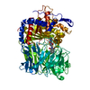 3eq7C  3eq9C  1qfsS C: citing same article ( S: Starting model for refinement |
|---|---|
| Similar structure data |


-Links
 PDBj
PDBj
- Assembly
| Deposited unit | 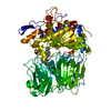
| ||||||||
|---|---|---|---|---|---|---|---|---|---|
| 1 |
| ||||||||
| Unit cell |
|
-Components
| #1: Protein | Mass: 80864.344 Da / Num. of mol.: 1 / Source method: isolated from a natural source / Source: (natural) |
|---|---|
| #2: Chemical | ChemComp-X98 /   Mass: 444.526 Da / Num. of mol.: 1 / Source method: obtained synthetically / Formula: C26H28N4O3 Mass: 444.526 Da / Num. of mol.: 1 / Source method: obtained synthetically / Formula: C26H28N4O3 |
| #3: Water | ChemComp-HOH /  Mass: 18.015 Da / Num. of mol.: 99 / Source method: isolated from a natural source / Formula: H2O Mass: 18.015 Da / Num. of mol.: 99 / Source method: isolated from a natural source / Formula: H2O |
-Experimental details
-Experiment
| Experiment | Method: X-RAY DIFFRACTION / Number of used crystals: 1 |
|---|
- Sample preparation
| Crystal | Density Matthews: 2.53 Å3/Da / Density % sol: 51.35 % |
|---|---|
| Crystal grow | Temperature: 277 K / Method: vapor diffusion, hanging drop / pH: 8.5 Details: 17% MPEG 5000, 15% glycerol, 20mM calcium acetate, 0.1M TRIS, pH 8.5, VAPOR DIFFUSION, HANGING DROP, temperature 277K |
-Data collection
| Diffraction | Mean temperature: 298 K |
|---|---|
| Diffraction source | Source: ROTATING ANODE / Type: RIGAKU RU200 / Wavelength: 1.5418 Å |
| Detector | Type: RIGAKU RAXIS IIC / Detector: IMAGE PLATE / Date: May 28, 1998 / Details: capillary collimator |
| Radiation | Monochromator: graphite / Protocol: SINGLE WAVELENGTH / Monochromatic (M) / Laue (L): M / Scattering type: x-ray |
| Radiation wavelength | Wavelength: 1.5418 Å / Relative weight: 1 |
| Reflection | Resolution: 2.724→34.378 Å / Num. obs: 21070 / % possible obs: 93.5 % / Observed criterion σ(F): 0 / Observed criterion σ(I): -3 / Redundancy: 4.2 % / Biso Wilson estimate: 47.11 Å2 / Rmerge(I) obs: 0.177 / Net I/σ(I): 3.9 |
| Reflection shell | Resolution: 2.724→2.88 Å / Redundancy: 2.1 % / Rmerge(I) obs: 0.521 / Num. unique all: 3047 / % possible all: 90.3 |
- Processing
| Software |
| ||||||||||||||||||||
|---|---|---|---|---|---|---|---|---|---|---|---|---|---|---|---|---|---|---|---|---|---|
| Refinement | Method to determine structure: FOURIER SYNTHESIS Starting model: PDB ENTRY 1QFS Resolution: 2.73→34.03 Å Isotropic thermal model: Grouped isotropic B-factors, 2 B-values/residue Cross valid method: THROUGHOUT / σ(F): 0 / Stereochemistry target values: Engh & Huber
| ||||||||||||||||||||
| Displacement parameters | Biso mean: 37.2 Å2 | ||||||||||||||||||||
| Refine analyze | Luzzati coordinate error obs: 0.2729 Å / Luzzati sigma a obs: 0.3156 Å | ||||||||||||||||||||
| Refinement step | Cycle: LAST / Resolution: 2.73→34.03 Å
| ||||||||||||||||||||
| Refine LS restraints |
|
