 ムービー
ムービー コントローラー


+ データを開く

- 基本情報
基本情報
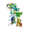
| 登録情報 | データベース: PDB / ID: 3e7o | ||||||
|---|---|---|---|---|---|---|---|
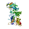
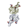
| タイトル | Crystal Structure of JNK2 | ||||||
 要素 要素 | Mitogen-activated protein kinase 9 | ||||||
 キーワード キーワード | TRANSFERASE / MAP kinase insert / activation loop / indazole inhibitor / ATP-binding / Kinase / Nucleotide-binding / Phosphoprotein / Serine/threonine-protein kinase | ||||||
| 機能・相同性 |  機能・相同性情報 機能・相同性情報protein localization to tricellular tight junction / JUN kinase activity / inflammatory response to wounding / positive regulation of macrophage derived foam cell differentiation / positive regulation of cytokine production involved in inflammatory response / positive regulation of podosome assembly / Activation of the AP-1 family of transcription factors / Fc-epsilon receptor signaling pathway / mitogen-activated protein kinase / JNK cascade ...protein localization to tricellular tight junction / JUN kinase activity / inflammatory response to wounding / positive regulation of macrophage derived foam cell differentiation / positive regulation of cytokine production involved in inflammatory response / positive regulation of podosome assembly / Activation of the AP-1 family of transcription factors / Fc-epsilon receptor signaling pathway / mitogen-activated protein kinase / JNK cascade / cellular response to cadmium ion / protein serine/threonine/tyrosine kinase activity / JNK (c-Jun kinases) phosphorylation and activation mediated by activated human TAK1 / positive regulation of protein ubiquitination / apoptotic signaling pathway / positive regulation of apoptotic signaling pathway / FCERI mediated MAPK activation / Schaffer collateral - CA1 synapse / modulation of chemical synaptic transmission / regulation of circadian rhythm / cellular response to reactive oxygen species / cellular senescence / Signaling by ALK fusions and activated point mutants / rhythmic process / positive regulation of proteasomal ubiquitin-dependent protein catabolic process / Oxidative Stress Induced Senescence / nuclear speck / protein phosphorylation / protein serine kinase activity / protein serine/threonine kinase activity / positive regulation of gene expression / mitochondrion / nucleoplasm / ATP binding / nucleus / plasma membrane / cytoplasm / cytosol 類似検索 - 分子機能 | ||||||
| 生物種 |  Homo sapiens (ヒト) Homo sapiens (ヒト) | ||||||
| 手法 |  X線回折 / シンクロトロン / 分子置換 / 解像度: 2.14 Å X線回折 / シンクロトロン / 分子置換 / 解像度: 2.14 Å | ||||||
 データ登録者 データ登録者 | Kuglstatter, A. / Villasenor, A.G. | ||||||
 引用 引用 | ジャーナル: J.Mol.Biol. / 年: 2008 タイトル: The crystal structure of JNK2 reveals conformational flexibility in the MAP kinase insert and indicates its involvement in the regulation of catalytic activity. 著者: Shaw, D. / Wang, S.M. / Villasenor, A.G. / Tsing, S. / Walter, D. / Browner, M.F. / Barnett, J. / Kuglstatter, A. | ||||||
| 履歴 |
|
- 構造の表示
構造の表示
| 構造ビューア | 分子: MolmilJmol/JSmol |
|---|
- ダウンロードとリンク
-ダウンロード
| PDBx/mmCIF形式 | 3e7o.cif.gz | 150.2 KB | 表示 | PDBx/mmCIF形式 |
|---|---|---|---|---|
| PDB形式 | pdb3e7o.ent.gz | 118.9 KB | 表示 | PDB形式 |
| PDBx/mmJSON形式 | 3e7o.json.gz | ツリー表示 | PDBx/mmJSON形式 | |
| その他 |  その他のダウンロード その他のダウンロード |
-検証レポート
| 文書・要旨 | 3e7o_validation.pdf.gz | 964.9 KB | 表示 | wwPDB検証レポート |
|---|---|---|---|---|
| 文書・詳細版 | 3e7o_full_validation.pdf.gz | 971.6 KB | 表示 | |
| XML形式データ | 3e7o_validation.xml.gz | 27.3 KB | 表示 | |
| CIF形式データ | 3e7o_validation.cif.gz | 38.7 KB | 表示 | |
| アーカイブディレクトリ | https://data.pdbj.org/pub/pdb/validation_reports/e7/3e7oftp://data.pdbj.org/pub/pdb/validation_reports/e7/3e7o | HTTPS FTP |
-関連構造データ
| 関連構造データ |  1jnkS S: 精密化の開始モデル |
|---|---|
| 類似構造データ |


-リンク
 PDBj
PDBj


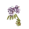
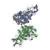
- 集合体
集合体
| 登録構造単位 | 
| ||||||||
|---|---|---|---|---|---|---|---|---|---|
| 1 | 
| ||||||||
| 2 | 
| ||||||||
| 3 | 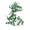
| ||||||||
| 単位格子 |
| ||||||||
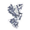
| 詳細 | The asymmetric unit contains two biological units which differ in conformation. |
-要素
| #1: タンパク質 | 分子量: 41271.430 Da / 分子数: 2 / 断片: residues 7-362 / 変異: C177S, C222S, K250A, K251A, K265A, K270A / 由来タイプ: 組換発現 / 由来: (組換発現) Homo sapiens (ヒト) / 遺伝子: MAPK9, JNK2, PRKM9 / プラスミド: pET15b / 発現宿主:  #2: 化合物 |   分子量: 370.364 Da / 分子数: 2 / 由来タイプ: 合成 / 式: C20H14N6O2 分子量: 370.364 Da / 分子数: 2 / 由来タイプ: 合成 / 式: C20H14N6O2#3: 水 | ChemComp-HOH / |  分子量: 18.015 Da / 分子数: 251 / 由来タイプ: 天然 / 式: H2O 分子量: 18.015 Da / 分子数: 251 / 由来タイプ: 天然 / 式: H2O |
|---|
-実験情報
-実験
| 実験 | 手法: X線回折 / 使用した結晶の数: 1 |
|---|
- 試料調製
試料調製
| 結晶 | マシュー密度: 3.03 Å3/Da / 溶媒含有率: 59.35 % |
|---|---|
| 結晶化 | 温度: 296 K / 手法: 蒸気拡散法, ハンギングドロップ法 / pH: 5.6 詳細: 0.1M MES, 0.6M sodium citrate, pH 5.6, VAPOR DIFFUSION, HANGING DROP, temperature 296K |
-データ収集
| 回折 | 平均測定温度: 100 K |
|---|---|
| 放射光源 | 由来: シンクロトロン / サイト: ALS  / ビームライン: 5.0.2 / 波長: 1 Å / ビームライン: 5.0.2 / 波長: 1 Å |
| 検出器 | タイプ: ADSC QUANTUM 315 / 検出器: CCD |
| 放射 | プロトコル: SINGLE WAVELENGTH / 単色(M)・ラウエ(L): M / 散乱光タイプ: x-ray |
| 放射波長 | 波長: 1 Å / 相対比: 1 |
| 反射 | 解像度: 2.14→50 Å / Num. all: 53458 / Num. obs: 51687 / % possible obs: 96.7 % / 冗長度: 2.7 % / Biso Wilson estimate: 40.6 Å2 / Rmerge(I) obs: 0.111 / Rsym value: 0.111 / Net I/σ(I): 8.4 |
| 反射 シェル | 解像度: 2.14→2.23 Å / 冗長度: 2.1 % / Rmerge(I) obs: 0.468 / Mean I/σ(I) obs: 1.9 / Num. unique all: 4473 / Rsym value: 0.468 / % possible all: 84 |
- 解析
解析
| ソフトウェア |
| ||||||||||||||||||||||||||||||||||||||||||||||||||||||||||||||||||||||||||||||||||||||||||
|---|---|---|---|---|---|---|---|---|---|---|---|---|---|---|---|---|---|---|---|---|---|---|---|---|---|---|---|---|---|---|---|---|---|---|---|---|---|---|---|---|---|---|---|---|---|---|---|---|---|---|---|---|---|---|---|---|---|---|---|---|---|---|---|---|---|---|---|---|---|---|---|---|---|---|---|---|---|---|---|---|---|---|---|---|---|---|---|---|---|---|---|
| 精密化 | 構造決定の手法: 分子置換 開始モデル: PDB ENTRY 1JNK 解像度: 2.14→44.29 Å / Cor.coef. Fo:Fc: 0.947 / Cor.coef. Fo:Fc free: 0.926 / SU B: 5.335 / SU ML: 0.14 / Isotropic thermal model: Isotropic / 交差検証法: THROUGHOUT / ESU R: 0.224 / ESU R Free: 0.189 / 立体化学のターゲット値: MAXIMUM LIKELIHOOD
| ||||||||||||||||||||||||||||||||||||||||||||||||||||||||||||||||||||||||||||||||||||||||||
| 溶媒の処理 | イオンプローブ半径: 0.8 Å / 減衰半径: 0.8 Å / VDWプローブ半径: 1.4 Å / 溶媒モデル: MASK | ||||||||||||||||||||||||||||||||||||||||||||||||||||||||||||||||||||||||||||||||||||||||||
| 原子変位パラメータ | Biso mean: 43.889 Å2
| ||||||||||||||||||||||||||||||||||||||||||||||||||||||||||||||||||||||||||||||||||||||||||
| 精密化ステップ | サイクル: LAST / 解像度: 2.14→44.29 Å
| ||||||||||||||||||||||||||||||||||||||||||||||||||||||||||||||||||||||||||||||||||||||||||
| 拘束条件 |
| ||||||||||||||||||||||||||||||||||||||||||||||||||||||||||||||||||||||||||||||||||||||||||
| LS精密化 シェル | 解像度: 2.143→2.199 Å / Total num. of bins used: 20
|
