protein localization to tricellular tight junction / JUN kinase activity / inflammatory response to wounding / positive regulation of macrophage derived foam cell differentiation / positive regulation of cytokine production involved in inflammatory response / positive regulation of podosome assembly / Activation of the AP-1 family of transcription factors / Fc-epsilon receptor signaling pathway / mitogen-activated protein kinase / response to cadmium ion ...protein localization to tricellular tight junction / JUN kinase activity / inflammatory response to wounding / positive regulation of macrophage derived foam cell differentiation / positive regulation of cytokine production involved in inflammatory response / positive regulation of podosome assembly / Activation of the AP-1 family of transcription factors / Fc-epsilon receptor signaling pathway / mitogen-activated protein kinase / response to cadmium ion / JNK cascade / protein serine/threonine/tyrosine kinase activity / positive regulation of protein ubiquitination / JNK (c-Jun kinases) phosphorylation and activation mediated by activated human TAK1 / positive regulation of apoptotic signaling pathway / FCERI mediated MAPK activation / cellular response to reactive oxygen species / apoptotic signaling pathway / regulation of circadian rhythm / modulation of chemical synaptic transmission / Schaffer collateral - CA1 synapse / cellular senescence / Signaling by ALK fusions and activated point mutants / rhythmic process / positive regulation of proteasomal ubiquitin-dependent protein catabolic process / Oxidative Stress Induced Senescence / protein phosphorylation / nuclear speck / protein serine kinase activity / protein serine/threonine kinase activity / positive regulation of gene expression / mitochondrion / nucleoplasm / ATP binding / nucleus / plasma membrane / cytoplasm / cytosol Similarity search - Function
Mitogen-activated protein (MAP) kinase, JNK / Mitogen-activated protein (MAP) kinase, conserved site / MAP kinase signature. / : / Phosphorylase Kinase; domain 1 / Phosphorylase Kinase; domain 1 / Transferase(Phosphotransferase) domain 1 / Transferase(Phosphotransferase); domain 1 / Serine/threonine-protein kinase, active site / Serine/Threonine protein kinases active-site signature. ...Mitogen-activated protein (MAP) kinase, JNK / Mitogen-activated protein (MAP) kinase, conserved site / MAP kinase signature. / : / Phosphorylase Kinase; domain 1 / Phosphorylase Kinase; domain 1 / Transferase(Phosphotransferase) domain 1 / Transferase(Phosphotransferase); domain 1 / Serine/threonine-protein kinase, active site / Serine/Threonine protein kinases active-site signature. / Protein kinase domain / Serine/Threonine protein kinases, catalytic domain / Protein kinase domain profile. / Protein kinase domain / Protein kinase-like domain superfamily / 2-Layer Sandwich / Orthogonal Bundle / Mainly Alpha / Alpha Beta Similarity search - Domain/homology
In the structure databanks used in Yorodumi, some data are registered as the other names, "COVID-19 virus" and "2019-nCoV". Here are the details of the virus and the list of structure data.
Jan 31, 2019. EMDB accession codes are about to change! (news from PDBe EMDB page)
EMDB accession codes are about to change! (news from PDBe EMDB page)
The allocation of 4 digits for EMDB accession codes will soon come to an end. Whilst these codes will remain in use, new EMDB accession codes will include an additional digit and will expand incrementally as the available range of codes is exhausted. The current 4-digit format prefixed with “EMD-” (i.e. EMD-XXXX) will advance to a 5-digit format (i.e. EMD-XXXXX), and so on. It is currently estimated that the 4-digit codes will be depleted around Spring 2019, at which point the 5-digit format will come into force.
The EM Navigator/Yorodumi systems omit the EMD- prefix.
Related info.:Q: What is EMD? / ID/Accession-code notation in Yorodumi/EM Navigator
Yorodumi is a browser for structure data from EMDB, PDB, SASBDB, etc.
This page is also the successor to EM Navigator detail page, and also detail information page/front-end page for Omokage search.
The word "yorodu" (or yorozu) is an old Japanese word meaning "ten thousand". "mi" (miru) is to see.
Related info.:EMDB / PDB / SASBDB / Comparison of 3 databanks / Yorodumi Search / Aug 31, 2016. New EM Navigator & Yorodumi / Yorodumi Papers / Jmol/JSmol / Function and homology information / Changes in new EM Navigator and Yorodumi
 Movie
Movie Controller


 Open data
 Basic information
 Components
 Keywords
 Function and homology information
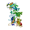
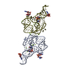
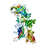
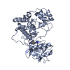
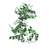
 Homo sapiens (human)
 X-RAY DIFFRACTION /
  Authors
 Citation
 Structure visualization
 Downloads & links
 Other downloads


 PDBj
PDBj


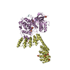
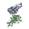
 Assembly


 Mass: 370.364 Da / Num. of mol.: 2 / Source method: obtained synthetically / Formula: C20H14N6O2
 Mass: 18.015 Da / Num. of mol.: 251 / Source method: isolated from a natural source / Formula: H2O
 Sample preparation
 / Beamline: 5.0.2 / Wavelength: 1 Å
 Processing