 Movie
Movie Controller

[English] 日本語
 Yorodumi
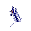
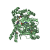
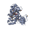
- PDB-3bss: PglD from Campylobacter jejuni, NCTC 11168, with native substrate -
+ Open data

- Basic information
| Entry | Database: PDB / ID: 3bss | ||||||
|---|---|---|---|---|---|---|---|
| Title | PglD from Campylobacter jejuni, NCTC 11168, with native substrate | ||||||
 Components Components | Acetyltransferase | ||||||
 Keywords Keywords | TRANSFERASE / left-hand beta helix / hexapeptide repeat / UDP / acetyl coenzyme Z / Rossmann fold / bacillosamine / campylobacter / pgl / N-linked glycosylation | ||||||
| Function / homology |  Function and homology information Function and homology informationUDP-N-acetylbacillosamine N-acetyltransferase / protein N-linked glycosylation via asparagine / acyltransferase activity, transferring groups other than amino-acyl groups Similarity search - Function | ||||||
| Biological species |   Campylobacter jejuni (Campylobacter) Campylobacter jejuni (Campylobacter) | ||||||
| Method |  X-RAY DIFFRACTION / SYNCHROTRON / MOLECULAR REPLACEMENT / molecular replacement / Resolution: 2.3 Å X-RAY DIFFRACTION / SYNCHROTRON / MOLECULAR REPLACEMENT / molecular replacement / Resolution: 2.3 Å | ||||||
 Authors Authors | Olivier, N.B. / Imperiali, B. | ||||||
 Citation Citation | Journal: J.Biol.Chem. / Year: 2008 Title: Crystal structure and catalytic mechanism of PglD from Campylobacter jejuni. Authors: Olivier, N.B. / Imperiali, B. | ||||||
| History |
|
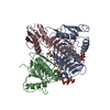
- Structure visualization
| Structure viewer | Molecule: MolmilJmol/JSmol |
|---|
- Downloads & links
-Download
| PDBx/mmCIF format | 3bss.cif.gz | 57.5 KB | Display | PDBx/mmCIF format |
|---|---|---|---|---|
| PDB format | pdb3bss.ent.gz | 39.9 KB | Display | PDB format |
| PDBx/mmJSON format | 3bss.json.gz | Tree view | PDBx/mmJSON format | |
| Others |  Other downloads Other downloads |
-Validation report
| Summary document | 3bss_validation.pdf.gz | 746.7 KB | Display | wwPDB validaton report |
|---|---|---|---|---|
| Full document | 3bss_full_validation.pdf.gz | 746.8 KB | Display | |
| Data in XML | 3bss_validation.xml.gz | 11.6 KB | Display | |
| Data in CIF | 3bss_validation.cif.gz | 16.8 KB | Display | |
| Arichive directory | https://data.pdbj.org/pub/pdb/validation_reports/bs/3bssftp://data.pdbj.org/pub/pdb/validation_reports/bs/3bss | HTTPS FTP |
-Related structure data
| Related structure data | 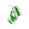 3bswSC  3bsyC S: Starting model for refinement C: citing same article ( |
|---|---|
| Similar structure data |


-Links
 PDBj
PDBj


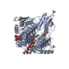
- Assembly
| Deposited unit | 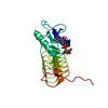
| ||||||||
|---|---|---|---|---|---|---|---|---|---|
| 1 | 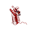
| ||||||||
| Unit cell |
| ||||||||
| Components on special symmetry positions |
|
-Components
| #1: Protein | Mass: 21389.957 Da / Num. of mol.: 1 Source method: isolated from a genetically manipulated source Source: (gene. exp.) Campylobacter jejuni (Campylobacter) / Strain: NCTC 11168 / Gene: pglDPlasmid details: N-terminal residues GSA are non-native; resulted from removal of N-terminal His-tag by thrombin Plasmid: pETGQ / Production host: References: UniProt: Q0P9D1, UDP-N-acetylglucosamine diphosphorylase |
|---|---|
| #2: Chemical | ChemComp-UD4 /   Mass: 590.370 Da / Num. of mol.: 1 / Source method: obtained synthetically / Formula: C17H28N4O15P2 Mass: 590.370 Da / Num. of mol.: 1 / Source method: obtained synthetically / Formula: C17H28N4O15P2 |
| #3: Water | ChemComp-HOH /  Mass: 18.015 Da / Num. of mol.: 202 / Source method: isolated from a natural source / Formula: H2O Mass: 18.015 Da / Num. of mol.: 202 / Source method: isolated from a natural source / Formula: H2O |
-Experimental details
-Experiment
| Experiment | Method: X-RAY DIFFRACTION / Number of used crystals: 1 |
|---|
- Sample preparation
| Crystal | Density Matthews: 8.3 Å3/Da / Density % sol: 85 % |
|---|---|
| Crystal grow | Temperature: 277 K / Method: vapor diffusion, hanging drop / pH: 6.5 Details: 1.3 M ammonium sulfate, 100 mM cacodylate, pH 6.5 in the resevior; protein solution containing 20 mM HEPES, 150 mM NaCl, pH 7.1, protein concentration of 10 mg/mL, UDP-4-amino-sugar at 5 mM; ...Details: 1.3 M ammonium sulfate, 100 mM cacodylate, pH 6.5 in the resevior; protein solution containing 20 mM HEPES, 150 mM NaCl, pH 7.1, protein concentration of 10 mg/mL, UDP-4-amino-sugar at 5 mM; drop made by mixing 1.5 uL of protein and resevoir solutions, VAPOR DIFFUSION, HANGING DROP, temperature 277K |
-Data collection
| Diffraction | Mean temperature: 110 K |
|---|---|
| Diffraction source | Source: SYNCHROTRON / Site: NSLS  / Beamline: X6A / Wavelength: 0.9784 Å / Beamline: X6A / Wavelength: 0.9784 Å |
| Detector | Type: ADSC QUANTUM 210 / Detector: CCD / Date: Oct 8, 2007 / Details: Toroidal focusing mirror |
| Radiation | Monochromator: Si(111) channel cut monochromator / Protocol: SINGLE WAVELENGTH / Monochromatic (M) / Laue (L): M / Scattering type: x-ray |
| Radiation wavelength | Wavelength: 0.9784 Å / Relative weight: 1 |
| Reflection | Resolution: 2.3→30 Å / Num. all: 33079 / Num. obs: 33079 / % possible obs: 100 % / Observed criterion σ(F): 0 / Observed criterion σ(I): 0 / Redundancy: 30.2 % / Biso Wilson estimate: 36.8 Å2 / Rmerge(I) obs: 0.11 / Χ2: 0.557 / Net I/σ(I): 5.8 |
| Reflection shell | Resolution: 2.3→2.38 Å / Redundancy: 30.8 % / Rmerge(I) obs: 0.607 / Mean I/σ(I) obs: 29.43 / Num. unique all: 3231 / Χ2: 0.507 / % possible all: 100 |
-Phasing
| Phasing | Method: molecular replacement | |||||||||
|---|---|---|---|---|---|---|---|---|---|---|
| Phasing MR |
|
- Processing
| Software |
| ||||||||||||||||||||||||||||||||||||||||||||||||||||||||||||||||||||||||||||||||||||||||||
|---|---|---|---|---|---|---|---|---|---|---|---|---|---|---|---|---|---|---|---|---|---|---|---|---|---|---|---|---|---|---|---|---|---|---|---|---|---|---|---|---|---|---|---|---|---|---|---|---|---|---|---|---|---|---|---|---|---|---|---|---|---|---|---|---|---|---|---|---|---|---|---|---|---|---|---|---|---|---|---|---|---|---|---|---|---|---|---|---|---|---|---|
| Refinement | Method to determine structure: MOLECULAR REPLACEMENT Starting model: PDB entry 3BSW Resolution: 2.3→29.64 Å / Cor.coef. Fo:Fc: 0.955 / Cor.coef. Fo:Fc free: 0.949 / Cross valid method: THROUGHOUT / σ(F): 0 / σ(I): 0 / ESU R: 0.109 / ESU R Free: 0.104 / Stereochemistry target values: MAXIMUM LIKELIHOOD
| ||||||||||||||||||||||||||||||||||||||||||||||||||||||||||||||||||||||||||||||||||||||||||
| Solvent computation | Ion probe radii: 0.8 Å / Shrinkage radii: 0.8 Å / VDW probe radii: 1.2 Å / Solvent model: MASK | ||||||||||||||||||||||||||||||||||||||||||||||||||||||||||||||||||||||||||||||||||||||||||
| Displacement parameters | Biso mean: 28.606 Å2 | ||||||||||||||||||||||||||||||||||||||||||||||||||||||||||||||||||||||||||||||||||||||||||
| Refinement step | Cycle: LAST / Resolution: 2.3→29.64 Å
| ||||||||||||||||||||||||||||||||||||||||||||||||||||||||||||||||||||||||||||||||||||||||||
| Refine LS restraints |
| ||||||||||||||||||||||||||||||||||||||||||||||||||||||||||||||||||||||||||||||||||||||||||
| LS refinement shell | Resolution: 2.3→2.36 Å / Total num. of bins used: 20
|
