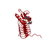
 Movie
Movie Controller


+ Open data

- Basic information
| Entry | Database: PDB / ID: 3bsw | ||||||
|---|---|---|---|---|---|---|---|
| Title | PglD-citrate complex, from Campylobacter jejuni NCTC 11168 | ||||||
 Components Components | Acetyltransferase | ||||||
 Keywords Keywords | TRANSFERASE / left-hand beta helix / hexapeptide repeat / UDP / acetyl coenzyme Z / Rossmann fold / bacillosamine / campylobacter / pgl / N-linked glycosylation | ||||||
| Function / homology |  Function and homology information Function and homology informationUDP-N-acetylbacillosamine N-acetyltransferase / : / acyltransferase activity, transferring groups other than amino-acyl groups Similarity search - Function | ||||||
| Biological species |   Campylobacter jejuni (Campylobacter) Campylobacter jejuni (Campylobacter) | ||||||
| Method |  X-RAY DIFFRACTION / SYNCHROTRON / SIRAS / Resolution: 1.77 Å X-RAY DIFFRACTION / SYNCHROTRON / SIRAS / Resolution: 1.77 Å | ||||||
| Model details | PglD with native substrate from Campylobacter jejuni, NCTC 11168 | ||||||
 Authors Authors | Olivier, N.B. / Imperiali, B. | ||||||
 Citation Citation | Journal: J.Biol.Chem. / Year: 2008 Title: Crystal structure and catalytic mechanism of PglD from Campylobacter jejuni. Authors: Olivier, N.B. / Imperiali, B. | ||||||
| History |
|
- Structure visualization
| Structure viewer | Molecule: MolmilJmol/JSmol |
|---|
- Downloads & links
-Download
| PDBx/mmCIF format | 3bsw.cif.gz | 54.7 KB | Display | PDBx/mmCIF format |
|---|---|---|---|---|
| PDB format | pdb3bsw.ent.gz | 38.3 KB | Display | PDB format |
| PDBx/mmJSON format | 3bsw.json.gz | Tree view | PDBx/mmJSON format | |
| Others |  Other downloads Other downloads |
-Validation report
| Arichive directory | https://data.pdbj.org/pub/pdb/validation_reports/bs/3bswftp://data.pdbj.org/pub/pdb/validation_reports/bs/3bsw | HTTPS FTP |
|---|
-Related structure data


-Links
 PDBj
PDBj


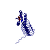
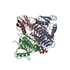
- Assembly
| Deposited unit | 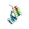
| ||||||||||||||||||
|---|---|---|---|---|---|---|---|---|---|---|---|---|---|---|---|---|---|---|---|
| 1 | 
| ||||||||||||||||||
| Unit cell |
| ||||||||||||||||||
| Components on special symmetry positions |
| ||||||||||||||||||
| Details | biological unit is a trimer generated from the monomer asymmetric unit by the operations -x+y,-x,z and -y,x-y,z |
-Components
| #1: Protein | Mass: 21389.957 Da / Num. of mol.: 1 Source method: isolated from a genetically manipulated source Details: N-terminal residues GSA are non-native; resulted from removal of N-terminal His-tag by thrombin Source: (gene. exp.) Campylobacter jejuni (Campylobacter) / Strain: NCTC 11168 / Gene: pglD / Plasmid: pETGQ / Production host: References: UniProt: Q0P9D1, UDP-N-acetylglucosamine diphosphorylase |
|---|---|
| #2: Chemical | ChemComp-CIT /   Mass: 192.124 Da / Num. of mol.: 1 / Source method: obtained synthetically / Formula: C6H8O7 Mass: 192.124 Da / Num. of mol.: 1 / Source method: obtained synthetically / Formula: C6H8O7 |
| #3: Water | ChemComp-HOH /  Mass: 18.015 Da / Num. of mol.: 206 / Source method: isolated from a natural source / Formula: H2O Mass: 18.015 Da / Num. of mol.: 206 / Source method: isolated from a natural source / Formula: H2O |
-Experimental details
-Experiment
| Experiment | Method: X-RAY DIFFRACTION / Number of used crystals: 2 |
|---|
- Sample preparation
| Crystal |
| |||||||||||||||
|---|---|---|---|---|---|---|---|---|---|---|---|---|---|---|---|---|
| Crystal grow |
|
-Data collection
| Diffraction |
| |||||||||||||||||||||||||||||||||||||||||||||||||||||||||||||||||||||||||||||
|---|---|---|---|---|---|---|---|---|---|---|---|---|---|---|---|---|---|---|---|---|---|---|---|---|---|---|---|---|---|---|---|---|---|---|---|---|---|---|---|---|---|---|---|---|---|---|---|---|---|---|---|---|---|---|---|---|---|---|---|---|---|---|---|---|---|---|---|---|---|---|---|---|---|---|---|---|---|---|
| Diffraction source | Source: SYNCHROTRON / Site: NSLS  / Beamline: X6A / Wavelength: 0.9784 Å / Beamline: X6A / Wavelength: 0.9784 Å | |||||||||||||||||||||||||||||||||||||||||||||||||||||||||||||||||||||||||||||
| Detector | Type: ADSC QUANTUM 210 / Detector: CCD / Date: Feb 7, 2007 / Details: Toroidal focusing mirror | |||||||||||||||||||||||||||||||||||||||||||||||||||||||||||||||||||||||||||||
| Radiation | Monochromator: Si(111) channel cut monochromator / Protocol: SINGLE WAVELENGTH / Monochromatic (M) / Laue (L): M / Scattering type: x-ray | |||||||||||||||||||||||||||||||||||||||||||||||||||||||||||||||||||||||||||||
| Radiation wavelength | Wavelength: 0.9784 Å / Relative weight: 1 | |||||||||||||||||||||||||||||||||||||||||||||||||||||||||||||||||||||||||||||
| Reflection | Redundancy: 5.6 % / Av σ(I) over netI: 5 / Number: 80527 / Rmerge(I) obs: 0.136 / Χ2: 1.01 / D res high: 2.2 Å / D res low: 30 Å / Num. obs: 14285 / % possible obs: 100 | |||||||||||||||||||||||||||||||||||||||||||||||||||||||||||||||||||||||||||||
| Diffraction reflection shell |
| |||||||||||||||||||||||||||||||||||||||||||||||||||||||||||||||||||||||||||||
| Reflection | Resolution: 1.77→30 Å / Num. all: 27293 / Num. obs: 27129 / % possible obs: 99.4 % / Observed criterion σ(F): 0 / Observed criterion σ(I): 0 / Redundancy: 5.7 % / Biso Wilson estimate: 25.11 Å2 / Rmerge(I) obs: 0.062 / Net I/σ(I): 27.7 | |||||||||||||||||||||||||||||||||||||||||||||||||||||||||||||||||||||||||||||
| Reflection shell | Resolution: 1.77→1.83 Å / Redundancy: 5.7 % / Rmerge(I) obs: 0.571 / Mean I/σ(I) obs: 2.9 / Num. unique all: 2695 / % possible all: 99.6 |
-Phasing
| Phasing | Method: SIRAS |
|---|
- Processing
| Software |
| ||||||||||||||||||||||||||||||||||||||||||||||||||||||||||||||||||||||||||||||||||||||||||
|---|---|---|---|---|---|---|---|---|---|---|---|---|---|---|---|---|---|---|---|---|---|---|---|---|---|---|---|---|---|---|---|---|---|---|---|---|---|---|---|---|---|---|---|---|---|---|---|---|---|---|---|---|---|---|---|---|---|---|---|---|---|---|---|---|---|---|---|---|---|---|---|---|---|---|---|---|---|---|---|---|---|---|---|---|---|---|---|---|---|---|---|
| Refinement | Method to determine structure: SIRAS / Resolution: 1.77→30 Å / Cor.coef. Fo:Fc: 0.962 / Cor.coef. Fo:Fc free: 0.961 / Cross valid method: THROUGHOUT / σ(F): 0 / σ(I): 0 / ESU R: 0.095 / ESU R Free: 0.09 / Stereochemistry target values: MAXIMUM LIKELIHOOD Details: Heavy atom sites in the substructure were identified using SHELX-D with data collected at the Se peak wavelength and truncated to 2.5 . Three out of five possible selenium sites for a single ...Details: Heavy atom sites in the substructure were identified using SHELX-D with data collected at the Se peak wavelength and truncated to 2.5 . Three out of five possible selenium sites for a single molecule of PglD in the asymmetric were located; CC All/weak=17.15/11.12, PATFOM 18.32. Structure factors from the native data were merged with initial phases using CAD (Riso=12%); phase extension to 1.77 and density modification were carried out using SHELX-E; values for contrast, connectivity, mean mapCC, and pseudo-free CC were 1.07, 0.96, 0.94, and 80.09%, respectively. The initial model was built with ARP/wARP (Perrakis et al., 2001) using the automated tracing function and manual adjustments were made using COOT (Emsley and Cowtan, 2004) and O (Jones et al., 1991).
| ||||||||||||||||||||||||||||||||||||||||||||||||||||||||||||||||||||||||||||||||||||||||||
| Solvent computation | Ion probe radii: 0.8 Å / Shrinkage radii: 0.8 Å / VDW probe radii: 1.2 Å / Solvent model: MASK | ||||||||||||||||||||||||||||||||||||||||||||||||||||||||||||||||||||||||||||||||||||||||||
| Displacement parameters | Biso mean: 25.892 Å2
| ||||||||||||||||||||||||||||||||||||||||||||||||||||||||||||||||||||||||||||||||||||||||||
| Refinement step | Cycle: LAST / Resolution: 1.77→30 Å
| ||||||||||||||||||||||||||||||||||||||||||||||||||||||||||||||||||||||||||||||||||||||||||
| Refine LS restraints |
| ||||||||||||||||||||||||||||||||||||||||||||||||||||||||||||||||||||||||||||||||||||||||||
| LS refinement shell | Resolution: 1.77→1.816 Å / Total num. of bins used: 20
|
