 Movie
Movie Controller

[English] 日本語
 Yorodumi
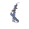
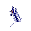
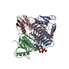
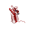
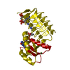
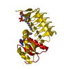
- PDB-2vhe: PglD-CoA complex: An acetyl transferase from Campylobacter jejuni -
+ Open data

- Basic information
| Entry | Database: PDB / ID: 2vhe | ||||||
|---|---|---|---|---|---|---|---|
| Title | PglD-CoA complex: An acetyl transferase from Campylobacter jejuni | ||||||
 Components Components | ACETYLTRANSFERASE | ||||||
 Keywords Keywords | TRANSFERASE / COENZYME A / N-ACETYL TRANSFERASE | ||||||
| Function / homology |  Function and homology information Function and homology informationUDP-N-acetylbacillosamine N-acetyltransferase / : / acyltransferase activity, transferring groups other than amino-acyl groups Similarity search - Function | ||||||
| Biological species |   CAMPYLOBACTER JEJUNI (Campylobacter) CAMPYLOBACTER JEJUNI (Campylobacter) | ||||||
| Method |  X-RAY DIFFRACTION / SYNCHROTRON / MOLECULAR REPLACEMENT / Resolution: 1.8 Å X-RAY DIFFRACTION / SYNCHROTRON / MOLECULAR REPLACEMENT / Resolution: 1.8 Å | ||||||
 Authors Authors | Rangarajan, E.S. / Ruane, K.M. / Sulea, T. / Watson, D.C. / Proteau, A. / Leclerc, S. / Cygler, M. / Matte, A. / Young, N.M. | ||||||
 Citation Citation | Journal: Biochemistry / Year: 2008 Title: Structure and Active Site Residues of Pgld, an N-Acetyltransferase from the Bacillosamine Synthetic Pathway Required for N-Glycan Synthesis in Campylobacter Jejuni Authors: Rangarajan, E.S. / Ruane, K.M. / Sulea, T. / Watson, D.C. / Proteau, A. / Leclerc, S. / Cygler, M. / Matte, A. / Young, N.M. | ||||||
| History |
|
- Structure visualization
| Structure viewer | Molecule: MolmilJmol/JSmol |
|---|
- Downloads & links
-Download
| PDBx/mmCIF format | 2vhe.cif.gz | 91.7 KB | Display | PDBx/mmCIF format |
|---|---|---|---|---|
| PDB format | pdb2vhe.ent.gz | 70.6 KB | Display | PDB format |
| PDBx/mmJSON format | 2vhe.json.gz | Tree view | PDBx/mmJSON format | |
| Others |  Other downloads Other downloads |
-Validation report
| Arichive directory | https://data.pdbj.org/pub/pdb/validation_reports/vh/2vheftp://data.pdbj.org/pub/pdb/validation_reports/vh/2vhe | HTTPS FTP |
|---|
-Related structure data
| Related structure data |  3bfpC  2npoS S: Starting model for refinement C: citing same article ( |
|---|---|
| Similar structure data |


-Links
 PDBj
PDBj


- Assembly
| Deposited unit | 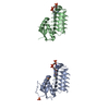
| |||||||||
|---|---|---|---|---|---|---|---|---|---|---|
| 1 | 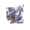
| |||||||||
| 2 | 
| |||||||||
| Unit cell |
| |||||||||
| Components on special symmetry positions |
| |||||||||
| Noncrystallographic symmetry (NCS) | NCS oper: (Code: given Matrix: (0.9907, -0.1361, -0.008404), Vector: |
-Components
| #1: Protein | Mass: 21043.553 Da / Num. of mol.: 2 / Fragment: RESIDUES 2-195 Source method: isolated from a genetically manipulated source Source: (gene. exp.) CAMPYLOBACTER JEJUNI (Campylobacter) / Production host: References: UniProt: Q0P9D1, UDP-N-acetylglucosamine diphosphorylase #2: Chemical |   Mass: 767.534 Da / Num. of mol.: 2 / Source method: obtained synthetically / Formula: C21H36N7O16P3S Mass: 767.534 Da / Num. of mol.: 2 / Source method: obtained synthetically / Formula: C21H36N7O16P3S#3: Chemical |   Mass: 96.063 Da / Num. of mol.: 2 / Source method: obtained synthetically / Formula: SO4 Mass: 96.063 Da / Num. of mol.: 2 / Source method: obtained synthetically / Formula: SO4#4: Water | ChemComp-HOH / |  Mass: 18.015 Da / Num. of mol.: 263 / Source method: isolated from a natural source / Formula: H2O Mass: 18.015 Da / Num. of mol.: 263 / Source method: isolated from a natural source / Formula: H2OSequence details | FIRST XXX CORESPOND TO THE TAG. NO. STARTS FROM ALA 2. EXPAND | |
|---|
-Experimental details
-Experiment
| Experiment | Method: X-RAY DIFFRACTION |
|---|
- Sample preparation
| Crystal | Density Matthews: 3.23 Å3/Da / Density % sol: 61.92 % / Description: NONE |
|---|---|
| Crystal grow | Details: 0.1 M SODIUM CITRATE PH 5.6, 0.16 M SODIUM TARTRATE AND 2.0 M AMMONIUM SULFATE |
-Data collection
| Diffraction | Mean temperature: 100 K |
|---|---|
| Diffraction source | Source: SYNCHROTRON / Site: ESRF  / Beamline: ID14-1 / Wavelength: 0.9756 / Beamline: ID14-1 / Wavelength: 0.9756 |
| Detector | Type: ADSC CCD / Detector: CCD / Date: Sep 8, 2005 |
| Radiation | Protocol: SINGLE WAVELENGTH / Monochromatic (M) / Laue (L): M / Scattering type: x-ray |
| Radiation wavelength | Wavelength: 0.9756 Å / Relative weight: 1 |
| Reflection | Resolution: 1.8→15 Å / Num. obs: 48692 / % possible obs: 100 % / Redundancy: 3.2 % / Rmerge(I) obs: 0.07 / Net I/σ(I): 11.4 |
| Reflection shell | Resolution: 1.8→1.9 Å / Redundancy: 3.2 % / Rmerge(I) obs: 0.46 / Mean I/σ(I) obs: 2.4 / % possible all: 100 |
- Processing
| Software |
| ||||||||||||||||||||||||||||||||||||||||||||||||||||||||||||||||||||||||||||||||||||||||||||||||||||||||||||||||||||||||||||||||||||||||||||||||||||||||||||||||||||||||||||||||||||||
|---|---|---|---|---|---|---|---|---|---|---|---|---|---|---|---|---|---|---|---|---|---|---|---|---|---|---|---|---|---|---|---|---|---|---|---|---|---|---|---|---|---|---|---|---|---|---|---|---|---|---|---|---|---|---|---|---|---|---|---|---|---|---|---|---|---|---|---|---|---|---|---|---|---|---|---|---|---|---|---|---|---|---|---|---|---|---|---|---|---|---|---|---|---|---|---|---|---|---|---|---|---|---|---|---|---|---|---|---|---|---|---|---|---|---|---|---|---|---|---|---|---|---|---|---|---|---|---|---|---|---|---|---|---|---|---|---|---|---|---|---|---|---|---|---|---|---|---|---|---|---|---|---|---|---|---|---|---|---|---|---|---|---|---|---|---|---|---|---|---|---|---|---|---|---|---|---|---|---|---|---|---|---|---|
| Refinement | Method to determine structure: MOLECULAR REPLACEMENT Starting model: PDB ENTRY 2NPO Resolution: 1.8→15 Å / Cor.coef. Fo:Fc: 0.957 / Cor.coef. Fo:Fc free: 0.945 / SU B: 4.977 / SU ML: 0.078 / TLS residual ADP flag: LIKELY RESIDUAL / Cross valid method: THROUGHOUT / ESU R: 0.108 / ESU R Free: 0.109 / Stereochemistry target values: MAXIMUM LIKELIHOOD Details: HYDROGENS HAVE BEEN ADDED IN THE RIDING POSITIONS. RESIDUES 36-46 AND 182-183 ARE DISORDERED
| ||||||||||||||||||||||||||||||||||||||||||||||||||||||||||||||||||||||||||||||||||||||||||||||||||||||||||||||||||||||||||||||||||||||||||||||||||||||||||||||||||||||||||||||||||||||
| Solvent computation | Ion probe radii: 0.8 Å / Shrinkage radii: 0.8 Å / VDW probe radii: 1.4 Å / Solvent model: MASK | ||||||||||||||||||||||||||||||||||||||||||||||||||||||||||||||||||||||||||||||||||||||||||||||||||||||||||||||||||||||||||||||||||||||||||||||||||||||||||||||||||||||||||||||||||||||
| Displacement parameters | Biso mean: 26.77 Å2
| ||||||||||||||||||||||||||||||||||||||||||||||||||||||||||||||||||||||||||||||||||||||||||||||||||||||||||||||||||||||||||||||||||||||||||||||||||||||||||||||||||||||||||||||||||||||
| Refinement step | Cycle: LAST / Resolution: 1.8→15 Å
| ||||||||||||||||||||||||||||||||||||||||||||||||||||||||||||||||||||||||||||||||||||||||||||||||||||||||||||||||||||||||||||||||||||||||||||||||||||||||||||||||||||||||||||||||||||||
| Refine LS restraints |
|