 Movie
Movie Controller

[English] 日本語
 Yorodumi
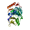
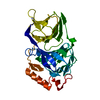
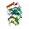
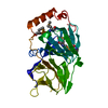
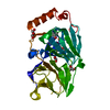
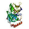
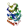
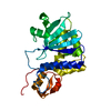
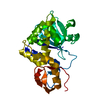
- PDB-3acl: Crystal Structure of Human Pirin in complex with Triphenyl Compound -
+ Open data

- Basic information
| Entry | Database: PDB / ID: 3acl | ||||||
|---|---|---|---|---|---|---|---|
| Title | Crystal Structure of Human Pirin in complex with Triphenyl Compound | ||||||
 Components Components | Pirin | ||||||
 Keywords Keywords | METAL BINDING PROTEIN / cupin / inhibitor / complex / Iron | ||||||
| Function / homology |  Function and homology information Function and homology informationDigestion / quercetin 2,3-dioxygenase / quercetin 2,3-dioxygenase activity / monocyte differentiation / digestion / transcription coregulator activity / transcription by RNA polymerase II / nuclear body / nucleoplasm / metal ion binding ...Digestion / quercetin 2,3-dioxygenase / quercetin 2,3-dioxygenase activity / monocyte differentiation / digestion / transcription coregulator activity / transcription by RNA polymerase II / nuclear body / nucleoplasm / metal ion binding / nucleus / cytosol / cytoplasm Similarity search - Function | ||||||
| Biological species |  Homo sapiens (human) Homo sapiens (human) | ||||||
| Method |  X-RAY DIFFRACTION / SYNCHROTRON / MOLECULAR REPLACEMENT / molecular replacement / Resolution: 2.35 Å X-RAY DIFFRACTION / SYNCHROTRON / MOLECULAR REPLACEMENT / molecular replacement / Resolution: 2.35 Å | ||||||
 Authors Authors | Okumura, H. / Miyazaki, I. / Simizu, S. / Osada, H. | ||||||
 Citation Citation | Journal: Nat.Chem.Biol. / Year: 2010 Title: A small-molecule inhibitor shows that pirin regulates migration of melanoma cells Authors: Miyazaki, I. / Simizu, S. / Okumura, H. / Takagi, S. / Osada, H. | ||||||
| History |
|
- Structure visualization
| Structure viewer | Molecule: MolmilJmol/JSmol |
|---|
- Downloads & links
-Download
| PDBx/mmCIF format | 3acl.cif.gz | 75.6 KB | Display | PDBx/mmCIF format |
|---|---|---|---|---|
| PDB format | pdb3acl.ent.gz | 53.2 KB | Display | PDB format |
| PDBx/mmJSON format | 3acl.json.gz | Tree view | PDBx/mmJSON format | |
| Others |  Other downloads Other downloads |
-Validation report
| Arichive directory | https://data.pdbj.org/pub/pdb/validation_reports/ac/3aclftp://data.pdbj.org/pub/pdb/validation_reports/ac/3acl | HTTPS FTP |
|---|
-Related structure data
| Related structure data |  1j1lS S: Starting model for refinement |
|---|---|
| Similar structure data |

-Links
 PDBj
PDBj


- Assembly
| Deposited unit | 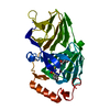
| ||||||||
|---|---|---|---|---|---|---|---|---|---|
| 1 |
| ||||||||
| Unit cell |
|
-Components
| #1: Protein | Mass: 32988.266 Da / Num. of mol.: 1 Source method: isolated from a genetically manipulated source Source: (gene. exp.) Homo sapiens (human) / Gene: PIR / Plasmid: pRSET C / Production host:  |
|---|---|
| #2: Chemical | ChemComp-FE2 /   Mass: 55.845 Da / Num. of mol.: 1 / Source method: obtained synthetically / Formula: Fe Mass: 55.845 Da / Num. of mol.: 1 / Source method: obtained synthetically / Formula: Fe |
| #3: Chemical | ChemComp-3F1 / 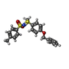  Mass: 399.526 Da / Num. of mol.: 1 / Source method: obtained synthetically / Formula: C21H21NO3S2 Mass: 399.526 Da / Num. of mol.: 1 / Source method: obtained synthetically / Formula: C21H21NO3S2 |
| #4: Water | ChemComp-HOH /  Mass: 18.015 Da / Num. of mol.: 131 / Source method: isolated from a natural source / Formula: H2O Mass: 18.015 Da / Num. of mol.: 131 / Source method: isolated from a natural source / Formula: H2O |
-Experimental details
-Experiment
| Experiment | Method: X-RAY DIFFRACTION / Number of used crystals: 1 |
|---|
- Sample preparation
| Crystal | Density Matthews: 2.33 Å3/Da / Density % sol: 47.25 % |
|---|---|
| Crystal grow | Temperature: 293 K / Method: vapor diffusion / pH: 6.5 Details: 34% PEG 3350, 0.1M PIPES, 0.1M MGCL2, 1.4mM INHIBITOR, PH 6.5, VAPOR DIFFUSION, TEMPERATURE 293K |
-Data collection
| Diffraction | Mean temperature: 100 K |
|---|---|
| Diffraction source | Source: SYNCHROTRON / Site: SPring-8  / Beamline: BL26B2 / Wavelength: 1 Å / Beamline: BL26B2 / Wavelength: 1 Å |
| Detector | Type: MARMOSAIC 225 mm CCD / Detector: CCD / Date: May 19, 2009 |
| Diffraction measurement | Details: 1.00 degrees, 31.2 sec, detector distance 200.00mm |
| Radiation | Monochromator: Si double crystal monochromator / Protocol: SINGLE WAVELENGTH / Monochromatic (M) / Laue (L): M / Scattering type: x-ray |
| Radiation wavelength | Wavelength: 1 Å / Relative weight: 1 |
| Reflection | Av R equivalents: 0.127 / Number: 13541 |
| Reflection | Resolution: 2.35→50 Å / Num. all: 13554 / Num. obs: 13541 / % possible obs: 99.9 % / Observed criterion σ(F): 0 / Observed criterion σ(I): 0 / Redundancy: 6.8 % / Biso Wilson estimate: 22.3 Å2 / Rmerge(I) obs: 0.127 / Rsym value: 0.127 / Net I/σ(I): 16 |
| Reflection shell | Resolution: 2.35→2.39 Å / Redundancy: 6.5 % / Rmerge(I) obs: 0.34 / Mean I/σ(I) obs: 3.803 / Rsym value: 0.34 / % possible all: 100 |
| Cell measurement | Reflection used: 13541 |
-Phasing
| Phasing | Method: molecular replacement | |||||||||
|---|---|---|---|---|---|---|---|---|---|---|
| Phasing MR |
|
- Processing
| Software |
| ||||||||||||||||||||||||||||||||||||
|---|---|---|---|---|---|---|---|---|---|---|---|---|---|---|---|---|---|---|---|---|---|---|---|---|---|---|---|---|---|---|---|---|---|---|---|---|---|
| Refinement | Method to determine structure: MOLECULAR REPLACEMENT Starting model: PDB ENTRY 1J1L Resolution: 2.35→35.86 Å / Rfactor Rfree error: 0.009 / Occupancy max: 1 / Occupancy min: 1 / Data cutoff high absF: 1297395 / Data cutoff low absF: 0 / Isotropic thermal model: Isotropic / Cross valid method: THROUGHOUT / σ(F): 0 / Stereochemistry target values: Engh & Huber / Details: BULK SOLVENT MODEL USED
| ||||||||||||||||||||||||||||||||||||
| Solvent computation | Solvent model: FLAT MODEL / Bsol: 19.223 Å2 / ksol: 0.35 e/Å3 | ||||||||||||||||||||||||||||||||||||
| Displacement parameters | Biso max: 50.33 Å2 / Biso mean: 19.79 Å2 / Biso min: 1.01 Å2
| ||||||||||||||||||||||||||||||||||||
| Refine analyze |
| ||||||||||||||||||||||||||||||||||||
| Refinement step | Cycle: LAST / Resolution: 2.35→35.86 Å
| ||||||||||||||||||||||||||||||||||||
| Refine LS restraints |
| ||||||||||||||||||||||||||||||||||||
| LS refinement shell | Resolution: 2.35→2.5 Å / Rfactor Rfree error: 0.031 / Total num. of bins used: 6
| ||||||||||||||||||||||||||||||||||||
| Xplor file |
|
