Movie
Movie Controller


+ Open data

- Basic information
| Entry | Database: EMDB / ID: EMD-3804 | |||||||||
|---|---|---|---|---|---|---|---|---|---|---|
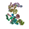
| Title | Cryo-EM structure of the yeast chromatin modifying complex SAGA | |||||||||
 Map data Map data | ||||||||||
 Sample Sample |
| |||||||||
| Biological species |  Komagataella pastoris (fungus) Komagataella pastoris (fungus) | |||||||||
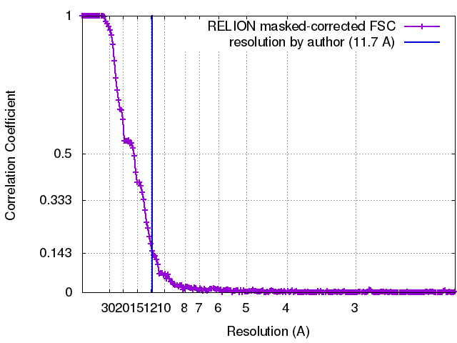
| Method | single particle reconstruction / cryo EM / Resolution: 11.7 Å | |||||||||
 Authors Authors | Sharov G / Voltz K / Durand A / Kolesnikova O / Dejaegere A / Papai G / Myasnikov AG / Ben-Shem A / Schultz P | |||||||||
 Citation Citation | Journal: Nat Commun / Year: 2017 Title: Structure of the transcription activator target Tra1 within the chromatin modifying complex SAGA. Authors: Grigory Sharov / Karine Voltz / Alexandre Durand / Olga Kolesnikova / Gabor Papai / Alexander G Myasnikov / Annick Dejaegere / Adam Ben Shem / Patrick Schultz /    Abstract: The transcription co-activator complex SAGA is recruited to gene promoters by sequence-specific transcriptional activators and by chromatin modifications to promote pre-initiation complex formation. ...The transcription co-activator complex SAGA is recruited to gene promoters by sequence-specific transcriptional activators and by chromatin modifications to promote pre-initiation complex formation. The yeast Tra1 subunit is the major target of acidic activators such as Gal4, VP16, or Gcn4 but little is known about its structural organization. The 430 kDa Tra1 subunit and its human homolog the transformation/transcription domain-associated protein TRRAP are members of the phosphatidyl 3-kinase-related kinase (PIKK) family. Here, we present the cryo-EM structure of the entire SAGA complex where the major target of activator binding, the 430 kDa Tra1 protein, is resolved with an average resolution of 5.7 Å. The high content of alpha-helices in Tra1 enabled tracing of the majority of its main chain. Our results highlight the integration of Tra1 within the major epigenetic regulator SAGA. | |||||||||
| History |
|
- Structure visualization
| Movie |
 Movie viewer Movie viewer |
|---|---|
| Structure viewer | EM map: SurfViewMolmilJmol/JSmol |
| Supplemental images |

- Downloads & links
-EMDB archive
| Map data | emd_3804.map.gz | 410.8 MB | EMDB map data format | |
|---|---|---|---|---|
| Header (meta data) | emd-3804-v30.xmlemd-3804.xml | 13.7 KB 13.7 KB | Display Display | EMDB header |
| FSC (resolution estimation) | emd_3804_fsc.xml | 17.9 KB | Display | FSC data file |
| Images | 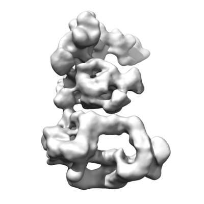 emd_3804.png emd_3804.png | 48 KB | ||
| Archive directory |  http://ftp.pdbj.org/pub/emdb/structures/EMD-3804ftp://ftp.pdbj.org/pub/emdb/structures/EMD-3804 http://ftp.pdbj.org/pub/emdb/structures/EMD-3804ftp://ftp.pdbj.org/pub/emdb/structures/EMD-3804 | HTTPS FTP |
-Validation report
| Summary document | emd_3804_validation.pdf.gz | 243.6 KB | Display | EMDB validaton report |
|---|---|---|---|---|
| Full document | emd_3804_full_validation.pdf.gz | 242.8 KB | Display | |
| Data in XML | emd_3804_validation.xml.gz | 16 KB | Display | |
| Arichive directory | https://ftp.pdbj.org/pub/emdb/validation_reports/EMD-3804ftp://ftp.pdbj.org/pub/emdb/validation_reports/EMD-3804 | HTTPS FTP |
-Related structure data


-Links
| EMDB pages | EMDB (EBI/PDBe) / EMDataResource |
|---|
-Map
| File | Download / File: emd_3804.map.gz / Format: CCP4 / Size: 512 MB / Type: IMAGE STORED AS FLOATING POINT NUMBER (4 BYTES) | ||||||||||||||||||||||||||||||||||||||||||||||||||||||||||||
|---|---|---|---|---|---|---|---|---|---|---|---|---|---|---|---|---|---|---|---|---|---|---|---|---|---|---|---|---|---|---|---|---|---|---|---|---|---|---|---|---|---|---|---|---|---|---|---|---|---|---|---|---|---|---|---|---|---|---|---|---|---|
| Voxel size | X=Y=Z: 1.1 Å | ||||||||||||||||||||||||||||||||||||||||||||||||||||||||||||
| Density |
| ||||||||||||||||||||||||||||||||||||||||||||||||||||||||||||
| Symmetry | Space group: 1 | ||||||||||||||||||||||||||||||||||||||||||||||||||||||||||||
| Details | EMDB XML:
CCP4 map header:
| ||||||||||||||||||||||||||||||||||||||||||||||||||||||||||||
-Supplemental data
- Sample components
-Entire : Spt-Ada-Gcn5-acetyltransferase complex (SAGA)
| Entire | Name: Spt-Ada-Gcn5-acetyltransferase complex (SAGA) |
|---|---|
| Components |
|
-Supramolecule #1: Spt-Ada-Gcn5-acetyltransferase complex (SAGA)
| Supramolecule | Name: Spt-Ada-Gcn5-acetyltransferase complex (SAGA) / type: complex / ID: 1 / Parent: 0 / Macromolecule list: all |
|---|---|
| Source (natural) | Organism: Komagataella pastoris (fungus) |
| Molecular weight | Theoretical: 1.8 MDa |
-Macromolecule #1: Cryo-EM structure of yeast SAGA complex
| Macromolecule | Name: Cryo-EM structure of yeast SAGA complex / type: protein_or_peptide / ID: 1 / Enantiomer: LEVO |
|---|---|
| Source (natural) | Organism: Komagataella pastoris (fungus) |
| Sequence | String: () |
-Experimental details
-Structure determination
| Method | cryo EM |
|---|---|
 Processing Processing | single particle reconstruction |
| Aggregation state | particle |
-Sample preparation
| Concentration | 0.2 mg/mL |
|---|---|
| Buffer | pH: 8 |
| Grid | Model: Quantifoil R2/2 / Material: COPPER / Mesh: 300 / Support film - Material: CARBON / Support film - topology: HOLEY / Pretreatment - Type: GLOW DISCHARGE / Pretreatment - Atmosphere: AIR / Pretreatment - Pressure: 0.018 kPa |
| Vitrification | Cryogen name: ETHANE / Chamber humidity: 95 % / Chamber temperature: 277 K / Instrument: FEI VITROBOT MARK IV / Details: Blot for 1 second before plunging. |
- Electron microscopy
| Microscope | FEI TITAN KRIOS |
|---|---|
| Temperature | Min: 70.0 K / Max: 80.0 K |
| Specialist optics | Spherical aberration corrector: Microscope has a Cs corrector |
| Image recording | Film or detector model: FEI FALCON II (4k x 4k) / Detector mode: INTEGRATING / Digitization - Dimensions - Width: 4096 pixel / Digitization - Dimensions - Height: 4096 pixel / Digitization - Sampling interval: 14.0 µm / Digitization - Frames/image: 2-8 / Number grids imaged: 4 / Number real images: 8505 / Average exposure time: 1.0 sec. / Average electron dose: 60.0 e/Å2 Details: Images were collected in movie-mode at 17 frames per second, frame 1 was not acquired. Every two frames were joined together, producing 8 frames per second. |
| Electron beam | Acceleration voltage: 300 kV / Electron source:  FIELD EMISSION GUN FIELD EMISSION GUN |
| Electron optics | C2 aperture diameter: 100.0 µm / Calibrated defocus max: 3.4 µm / Calibrated defocus min: 1.4 µm / Calibrated magnification: 127272 / Illumination mode: FLOOD BEAM / Imaging mode: BRIGHT FIELD / Cs: 0.001 mm / Nominal defocus max: 3.4 µm / Nominal defocus min: 1.4 µm / Nominal magnification: 59000 |
| Sample stage | Specimen holder model: FEI TITAN KRIOS AUTOGRID HOLDER / Cooling holder cryogen: NITROGEN |
| Experimental equipment |  Model: Titan Krios / Image courtesy: FEI Company |