 Movie
Movie Controller

[English] 日本語
 Yorodumi
- EMDB-3750: Cryo-EM reconstruction of the large subunit of the Mycobacterium ... -
+ Open data

- Basic information
| Entry | Database: EMDB / ID: EMD-3750 | |||||||||
|---|---|---|---|---|---|---|---|---|---|---|
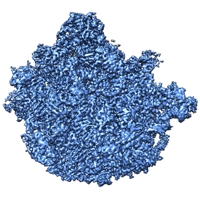
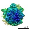
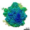
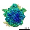
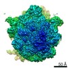
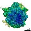
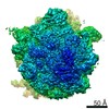
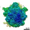
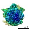
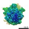
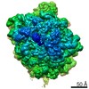
| Title | Cryo-EM reconstruction of the large subunit of the Mycobacterium smegmatis ribosome | |||||||||
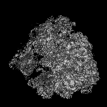
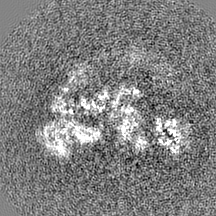
 Map data Map data | ||||||||||
 Sample Sample |
| |||||||||
 Keywords Keywords | ribosome / translation | |||||||||
| Function / homology |  Function and homology information Function and homology informationlarge ribosomal subunit / transferase activity / 5S rRNA binding / ribosomal large subunit assembly / large ribosomal subunit rRNA binding / cytosolic large ribosomal subunit / cytoplasmic translation / tRNA binding / negative regulation of translation / rRNA binding ...large ribosomal subunit / transferase activity / 5S rRNA binding / ribosomal large subunit assembly / large ribosomal subunit rRNA binding / cytosolic large ribosomal subunit / cytoplasmic translation / tRNA binding / negative regulation of translation / rRNA binding / structural constituent of ribosome / ribosome / translation / ribonucleoprotein complex / mRNA binding / metal ion binding / cytoplasm Similarity search - Function | |||||||||
| Biological species |  Mycobacterium smegmatis str. MC2 155 (bacteria) Mycobacterium smegmatis str. MC2 155 (bacteria) | |||||||||
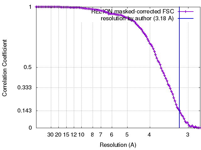
| Method | single particle reconstruction / cryo EM / Resolution: 3.18 Å | |||||||||
 Authors Authors | Hentschel J / Burnside C | |||||||||
| Funding support |  Switzerland, 2 items Switzerland, 2 items
| |||||||||
 Citation Citation | Journal: Cell Rep / Year: 2017 Title: The Complete Structure of the Mycobacterium smegmatis 70S Ribosome. Authors: Jendrik Hentschel / Chloe Burnside / Ingrid Mignot / Marc Leibundgut / Daniel Boehringer / Nenad Ban / Abstract: The ribosome carries out the synthesis of proteins in every living cell. It consequently represents a frontline target in anti-microbial therapy. Tuberculosis ranks among the leading causes of death ...The ribosome carries out the synthesis of proteins in every living cell. It consequently represents a frontline target in anti-microbial therapy. Tuberculosis ranks among the leading causes of death worldwide, due in large part to the combination of difficult-to-treat latency and antibiotic resistance. Here, we present the 3.3-Å cryo-EM structure of the 70S ribosome of Mycobacterium smegmatis, a close relative to the human pathogen Mycobacterium tuberculosis. The structure reveals two additional ribosomal proteins and localizes them to the vicinity of drug-target sites in both the catalytic center and the decoding site of the ribosome. Furthermore, we visualized actinobacterium-specific rRNA and protein expansions that extensively remodel the ribosomal surface with implications for polysome organization. Our results provide a foundation for understanding the idiosyncrasies of mycobacterial translation and reveal atomic details of the structure that will facilitate the design of anti-tubercular therapeutics. | |||||||||
| History |
|
- Structure visualization
| Movie |
Movie viewer |
|---|---|
| Structure viewer | EM map: SurfViewMolmilJmol/JSmol |
| Supplemental images |
- Downloads & links
-EMDB archive
| Map data | emd_3750.map.gz | 35.8 MB | EMDB map data format | |
|---|---|---|---|---|
| Header (meta data) | emd-3750-v30.xmlemd-3750.xml | 48.4 KB 48.4 KB | Display Display | EMDB header |
| FSC (resolution estimation) | emd_3750_fsc.xml | 10.1 KB | Display | FSC data file |
| Images |  emd_3750.png emd_3750.png | 231.5 KB | ||
| Filedesc metadata | emd-3750.cif.gz | 10.5 KB | ||
| Archive directory |  http://ftp.pdbj.org/pub/emdb/structures/EMD-3750ftp://ftp.pdbj.org/pub/emdb/structures/EMD-3750 http://ftp.pdbj.org/pub/emdb/structures/EMD-3750ftp://ftp.pdbj.org/pub/emdb/structures/EMD-3750 | HTTPS FTP |
-Related structure data
| Related structure data | 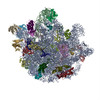 5o60MC 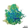 3748C 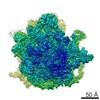 3751C 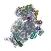 5o5jC 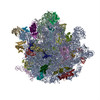 5o61C C: citing same article ( M: atomic model generated by this map |
|---|---|
| Similar structure data |
-Links
| EMDB pages | EMDB (EBI/PDBe) / EMDataResource |
|---|---|
| Related items in Molecule of the Month |


-Map
| File | Download / File: emd_3750.map.gz / Format: CCP4 / Size: 38.4 MB / Type: IMAGE STORED AS FLOATING POINT NUMBER (4 BYTES) | ||||||||||||||||||||||||||||||||||||||||||||||||||||||||||||
|---|---|---|---|---|---|---|---|---|---|---|---|---|---|---|---|---|---|---|---|---|---|---|---|---|---|---|---|---|---|---|---|---|---|---|---|---|---|---|---|---|---|---|---|---|---|---|---|---|---|---|---|---|---|---|---|---|---|---|---|---|---|
| Projections & slices | Image control
Images are generated by Spider. | ||||||||||||||||||||||||||||||||||||||||||||||||||||||||||||
| Voxel size | X=Y=Z: 1.39 Å | ||||||||||||||||||||||||||||||||||||||||||||||||||||||||||||
| Density |
| ||||||||||||||||||||||||||||||||||||||||||||||||||||||||||||
| Symmetry | Space group: 1 | ||||||||||||||||||||||||||||||||||||||||||||||||||||||||||||
| Details | EMDB XML:
CCP4 map header:
| ||||||||||||||||||||||||||||||||||||||||||||||||||||||||||||
 Z (Sec.)
 Y (Row.)
 X (Col.)


-Supplemental data
- Sample components
+Entire : 50S large ribosomal subunit
+Supramolecule #1: 50S large ribosomal subunit
+Macromolecule #1: 50S ribosomal protein bL37
+Macromolecule #4: 50S ribosomal protein L2
+Macromolecule #5: 50S ribosomal protein L3
+Macromolecule #6: 50S ribosomal protein L4
+Macromolecule #7: 50S ribosomal protein L5
+Macromolecule #8: 50S ribosomal protein L6
+Macromolecule #9: 50S ribosomal protein L9
+Macromolecule #10: 50S ribosomal protein L10
+Macromolecule #11: 50S ribosomal protein L11
+Macromolecule #12: 50S ribosomal protein L13
+Macromolecule #13: 50S ribosomal protein L14
+Macromolecule #14: 50S ribosomal protein L15
+Macromolecule #15: 50S ribosomal protein L16
+Macromolecule #16: 50S ribosomal protein L17
+Macromolecule #17: 50S ribosomal protein L18
+Macromolecule #18: 50S ribosomal protein L19
+Macromolecule #19: 50S ribosomal protein L20
+Macromolecule #20: 50S ribosomal protein L21
+Macromolecule #21: 50S ribosomal protein L22
+Macromolecule #22: 50S ribosomal protein L23
+Macromolecule #23: 50S ribosomal protein L24
+Macromolecule #24: 50S ribosomal protein L25
+Macromolecule #25: 50S ribosomal protein L27
+Macromolecule #26: 50S ribosomal protein L28
+Macromolecule #27: 50S ribosomal protein L29
+Macromolecule #28: 50S ribosomal protein L30
+Macromolecule #29: 50S ribosomal protein L32
+Macromolecule #30: 50S ribosomal protein L33 1
+Macromolecule #31: 50S ribosomal protein L34
+Macromolecule #32: 50S ribosomal protein L35
+Macromolecule #33: 50S ribosomal protein L36
+Macromolecule #34: 50S ribosomal protein L31
+Macromolecule #2: 23S rRNA
+Macromolecule #3: 5S rRNA
+Macromolecule #35: tRNA CCA-end acetylated (Phe)
+Macromolecule #36: MAGNESIUM ION
+Macromolecule #37: ZINC ION
+Macromolecule #38: PHENYLALANINE

-Experimental details
-Structure determination
| Method | cryo EM |
|---|---|
 Processing Processing | single particle reconstruction |
| Aggregation state | particle |
-Sample preparation
| Buffer | pH: 7.5 |
|---|---|
| Grid | Model: Quantifoil R2/2 / Material: COPPER |
| Vitrification | Cryogen name: ETHANE-PROPANE / Chamber humidity: 96 % / Instrument: FEI VITROBOT MARK I |
- Electron microscopy
| Microscope | FEI TITAN KRIOS |
|---|---|
| Image recording | Film or detector model: FEI FALCON II (4k x 4k) / Detector mode: INTEGRATING / Average electron dose: 20.0 e/Å2 / Details: FEI EPU data collection |
| Electron beam | Acceleration voltage: 300 kV / Electron source:  FIELD EMISSION GUN FIELD EMISSION GUN |
| Electron optics | Calibrated magnification: 100719 / Illumination mode: FLOOD BEAM / Imaging mode: BRIGHT FIELD |
| Sample stage | Specimen holder model: FEI TITAN KRIOS AUTOGRID HOLDER / Cooling holder cryogen: NITROGEN |
| Experimental equipment |  Model: Titan Krios / Image courtesy: FEI Company |
+Image processing
-Atomic model buiding 1
| Refinement | Protocol: OTHER |
|---|---|
| Output model | PDB-5o60: |
