 Movie
Movie Controller


+ Open data

- Basic information
| Entry | Database: EMDB / ID: EMD-3577 | |||||||||
|---|---|---|---|---|---|---|---|---|---|---|
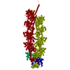
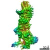
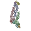
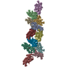
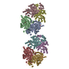
| Title | MTA2-RBBP7 complex | |||||||||
 Map data Map data | ||||||||||
 Sample Sample |
| |||||||||
| Biological species |  Homo sapiens (human) Homo sapiens (human) | |||||||||
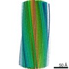
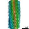
| Method | single particle reconstruction / negative staining / Resolution: 38.0 Å | |||||||||
 Authors Authors | Brasen C / Dorosz J / Wiuf A / Boesen T / Mirza O / Gajhede M | |||||||||
 Citation Citation | Journal: Biochim Biophys Acta Proteins Proteom / Year: 2017 Title: Expression, purification and characterization of the human MTA2-RBBP7 complex. Authors: Christoffer Brasen / Jerzy Dorosz / Anders Wiuf / Thomas Boesen / Osman Mirza / Michael Gajhede /  Abstract: The repressive Nucleosome Remodeling and histone Deacetylation (NuRD) complex remodels the chromatin structure by coupling ATP-dependent remodeling activity with histone deacetylase function and ...The repressive Nucleosome Remodeling and histone Deacetylation (NuRD) complex remodels the chromatin structure by coupling ATP-dependent remodeling activity with histone deacetylase function and plays important roles in regulating gene transcription, DNA damage repair and chromatin assembly. The complex is composed of six subunits: Metastasis Associated proteins MTA1/2/3 initially recruit histone chaperones RBBP4/7 followed by the histone deacetylases HDAC1/2 forming a core complex. Further association of the CpG-binding protein MBD2/3, p66α/β and the ATP-dependent helicase CDH3/4 constitutes the NuRD complex. Recent structural studies on truncated human proteins or orthologous have revealed that the stoichiometry of the MTA1-RBBP4 complex is 2:4. This study reports expression and purification of the intact human MTA2-RBBP7 complex using HEK293F cells as expression system. In analogy with findings on the Drosophila NuRD complex, we find that also the human MTA-RBBP can be isolated in vitro. Taken together with previous findings this suggests, that MTA-RBBP is a stable complex, with a central role in the initial assembly of the human NuRD complex. Refined 3D volumes of the complex generated from negative stain electron microscopy (EM) data reveals an elongated architecture that is capable of hinge like motion around the center of the particle. | |||||||||
| History |
|
- Structure visualization
| Movie |
 Movie viewer Movie viewer |
|---|---|
| Structure viewer | EM map: SurfViewMolmilJmol/JSmol |
| Supplemental images |
 UCSF Chimera
- Downloads & links
-EMDB archive
| Map data | emd_3577.map.gz | 13.5 MB | EMDB map data format | |
|---|---|---|---|---|
| Header (meta data) | emd-3577-v30.xmlemd-3577.xml | 9.5 KB 9.5 KB | Display Display | EMDB header |
| Images | 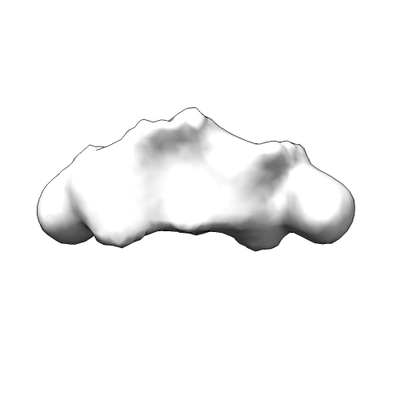 emd_3577.png emd_3577.png | 18.6 KB | ||
| Archive directory |  http://ftp.pdbj.org/pub/emdb/structures/EMD-3577ftp://ftp.pdbj.org/pub/emdb/structures/EMD-3577 http://ftp.pdbj.org/pub/emdb/structures/EMD-3577ftp://ftp.pdbj.org/pub/emdb/structures/EMD-3577 | HTTPS FTP |
-Related structure data
| Similar structure data |
|---|

-Links
| EMDB pages | EMDB (EBI/PDBe) / EMDataResource |
|---|
-Map
| File | Download / File: emd_3577.map.gz / Format: CCP4 / Size: 18.7 MB / Type: IMAGE STORED AS FLOATING POINT NUMBER (4 BYTES) | ||||||||||||||||||||||||||||||||||||||||||||||||||||||||||||
|---|---|---|---|---|---|---|---|---|---|---|---|---|---|---|---|---|---|---|---|---|---|---|---|---|---|---|---|---|---|---|---|---|---|---|---|---|---|---|---|---|---|---|---|---|---|---|---|---|---|---|---|---|---|---|---|---|---|---|---|---|---|
| Projections & slices | Image control
Images are generated by Spider. | ||||||||||||||||||||||||||||||||||||||||||||||||||||||||||||
| Voxel size | X=Y=Z: 3.15 Å | ||||||||||||||||||||||||||||||||||||||||||||||||||||||||||||
| Density |
| ||||||||||||||||||||||||||||||||||||||||||||||||||||||||||||
| Symmetry | Space group: 1 | ||||||||||||||||||||||||||||||||||||||||||||||||||||||||||||
| Details | EMDB XML:
CCP4 map header:
| ||||||||||||||||||||||||||||||||||||||||||||||||||||||||||||
 Z (Sec.)
 Y (Row.)
 X (Col.)


-Supplemental data
- Sample components
-Entire : MTA2-RBBP7 complex
| Entire | Name: MTA2-RBBP7 complex |
|---|---|
| Components |
|
-Supramolecule #1: MTA2-RBBP7 complex
| Supramolecule | Name: MTA2-RBBP7 complex / type: complex / ID: 1 / Parent: 0 / Macromolecule list: all |
|---|---|
| Source (natural) | Organism: Homo sapiens (human) |
| Recombinant expression | Organism: Homo sapiens (human) / Recombinant cell: HEK293F / Recombinant plasmid: pOPINHALO7/pOPINF |
-Macromolecule #1: MTA2-RBBP7 complex
| Macromolecule | Name: MTA2-RBBP7 complex / type: other / ID: 1 / Classification: other |
|---|---|
| Source (natural) | Organism: Homo sapiens (human) |
| Sequence | String: MAANMYRVGD YVYFENSSSN PYLVRRIEEL NKTANGNVEA KVVCLFRRRD ISSSLNSLAD SNAREFEEE SKQPGVSEQQ RHQLKHRELF LSRQFESLPA THIRGKCSVT LLNETDILSQ Y LEKEDCFF YSLVFDPVQK TLLADQGEIR VGCKYQAEIP DRLVEGESDN ...String: MAANMYRVGD YVYFENSSSN PYLVRRIEEL NKTANGNVEA KVVCLFRRRD ISSSLNSLAD SNAREFEEE SKQPGVSEQQ RHQLKHRELF LSRQFESLPA THIRGKCSVT LLNETDILSQ Y LEKEDCFF YSLVFDPVQK TLLADQGEIR VGCKYQAEIP DRLVEGESDN RNQQKMEMKV WD PDNPLTD RQIDQFLVVA RAVGTFARAL DCSSSIRQPS LHMSAAAASR DITLFHAMDT LQR NGYDLA KAMSTLVPQG GPVLCRDEME EWSASEAMLF EEALEKYGKD FNDIRQDFLP WKSL ASIVQ FYYMWKTTDR YIQQKRLKAA EADSKLKQVY IPTYTKPNPN QIISVGSKPG MNGAG FQKG LTCESCHTTQ SAQWYAWGPP NMQCRLCASC WIYWKKYGGL KTPTQLEGAT RGTTEP HSR GHLSRPEAQS LSPYTTSANR AKLLAKNRQT FLLQTTKLTR LARRMCRDLL QPRRAAR RP YAPINANAIK AECSIRLPKA AKTPLKIHPL VRLPLATIVK DLVAQAPLKP KTPRGTKT P INRNQLSQNR GLGGIMVKRA YETMAGAGVP FSANGRPLAS GIRSSSQPAA KRQKLNPAD APNPVVFVAT KDTRALRKAL THLEMRRAAR RPNLPLKVKP TLIAVRPPVP LPAPSHPAST NEPIVLED MASKEMFEDT VEERVINEEY KIWKKNTPFL YDLVMTHALQ WPSLTVQWLP EVTKPEGKDY ALHWLVLGT HTSDEQNHLV VARVHIPNDD AQFDASHCDS DKGEFGGFGS VTGKIECEIK I NHEGEVNR ARYMPQNPHI IATKTPSSDV LVFDYTKHPA KPDPSGECNP DLRLRGHQKE GY GLSWNSN LSGHLLSASD DHTVCLWDIN AGPKEGKIVD AKAIFTGHSA VVEDVAWHLL HES LFGSVA DDQKLMIWDT RSNTTSKPSH LVDAHTAEVN CLSFNPYSEF ILATGSADKT VALW DLRNL KLKLHTFESH KDEIFQVHWS PHNETILASS GTDRRLNVWD LSKIGEEQSA EDAED GPPE LLFIHGGHTA KISDFSWNPN EPWVICSVSE DNIMQIWQMA ENIYNDEESD VTTSEL EGQ GS |
| Recombinant expression | Organism: Homo sapiens (human) |
-Experimental details
-Structure determination
| Method | negative staining |
|---|---|
 Processing Processing | single particle reconstruction |
| Aggregation state | particle |
-Sample preparation
| Buffer | pH: 7.5 |
|---|---|
| Staining | Type: NEGATIVE / Material: uranyl formate |
- Electron microscopy
| Microscope | FEI TECNAI SPIRIT |
|---|---|
| Image recording | Film or detector model: TVIPS TEMCAM-F416 (4k x 4k) / Average electron dose: 10.0 e/Å2 |
| Electron beam | Acceleration voltage: 120 kV / Electron source: LAB6 |
| Electron optics | C2 aperture diameter: 100.0 µm / Illumination mode: OTHER / Imaging mode: BRIGHT FIELD / Cs: 2.2 mm |
| Experimental equipment |  Model: Tecnai Spirit / Image courtesy: FEI Company |
-Image processing
| Final reconstruction | Resolution.type: BY AUTHOR / Resolution: 38.0 Å / Resolution method: FSC 0.143 CUT-OFF / Number images used: 1267 |
|---|---|
| Initial angle assignment | Type: NOT APPLICABLE |
| Final angle assignment | Type: NOT APPLICABLE |
