[English] 日本語
Yorodumi
- EMDB-32192: STRUCTURE OF PHOTOSYNTHETIC LH1-RC SUPER-COMPLEX OF RHODOBACTER S... -

+
Open data


ID or keywords:

Loading...

-
Basic information

Entry
Database: EMDB / ID: EMD-32192
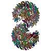
TitleSTRUCTURE OF PHOTOSYNTHETIC LH1-RC SUPER-COMPLEX OF RHODOBACTER SPHAEROIDES DIMER
Map data
Sample
  • Complex: Photosynthetic LH1-RC complex from the purple phototrophic bacterium Rhodobacter sphaeroides dimer
    • Complex: Rhodobacter sphaeroides Dimeric LH1-RC Complex
      • Protein or peptide: x 7 types
  • Ligand: x 10 types
KeywordsLH1-RC COMPLEX / PHOTOSYNTHESIS / PURPLE BACTERIA
Function / homology
Function and homology information


organelle inner membrane / plasma membrane-derived chromatophore membrane / plasma membrane light-harvesting complex / bacteriochlorophyll binding / photosynthesis, light reaction / photosynthetic electron transport in photosystem II / electron transporter, transferring electrons within the cyclic electron transport pathway of photosynthesis activity / membrane => GO:0016020 / metal ion binding / plasma membrane
Similarity search - Function
Intrinsic membrane protein family, PufX / Intrinsic membrane protein PufX / Antenna complex, beta subunit, conserved site / Antenna complexes beta subunits signature. / Antenna complex, alpha subunit / Antenna complex, alpha subunit conserved site / Antenna complexes alpha subunits signature. / Antenna complex, alpha/beta subunit / Light-harvesting protein B beta chain / Antenna complex, beta domain superfamily ...Intrinsic membrane protein family, PufX / Intrinsic membrane protein PufX / Antenna complex, beta subunit, conserved site / Antenna complexes beta subunits signature. / Antenna complex, alpha subunit / Antenna complex, alpha subunit conserved site / Antenna complexes alpha subunits signature. / Antenna complex, alpha/beta subunit / Light-harvesting protein B beta chain / Antenna complex, beta domain superfamily / Antenna complex alpha/beta subunit / Light-harvesting complex / Photosynthetic reaction centre, H subunit / Bacterial photosynthetic reaction centre, H-chain, C-terminal / Photosynthetic reaction centre, M subunit / Photosynthetic reaction centre, H subunit, N-terminal / Photosynthetic reaction centre, H subunit, N-terminal domain superfamily / Photosynthetic reaction centre, H-chain N-terminal region / PRC-barrel domain / PRC-barrel domain / Photosynthetic reaction centre, L subunit / PRC-barrel-like superfamily / Photosynthetic reaction centre, L/M / Photosystem II protein D1/D2 superfamily / Photosynthetic reaction centre protein / Photosynthetic reaction center proteins signature.
Similarity search - Domain/homology
Uncharacterized protein / Intrinsic membrane protein PufX / Reaction center protein L chain / Antenna pigment protein beta chain / Reaction center protein M chain / Photosynthetic reaction center subunit H / Antenna pigment protein alpha chain
Similarity search - Component
Biological speciesRhodobacter sphaeroides f. sp. denitrificans (bacteria)
Methodsingle particle reconstruction / cryo EM / Resolution: 2.75 Å
AuthorsTani K / Kanno R
Funding support Japan, 6 items
OrganizationGrant numberCountry
Japan Agency for Medical Research and Development (AMED)JP21am0101118 Japan
Japan Agency for Medical Research and Development (AMED)JP21am0101116 Japan
Japan Society for the Promotion of Science (JSPS)JP16H04174 Japan
Japan Society for the Promotion of Science (JSPS)JP18H05153 Japan
Japan Society for the Promotion of Science (JSPS)20H05086 Japan
Japan Society for the Promotion of Science (JSPS)20H02856 Japan
CitationJournal: Nat Commun / Year: 2022
Title: Asymmetric structure of the native Rhodobacter sphaeroides dimeric LH1-RC complex.
Authors: Kazutoshi Tani / Ryo Kanno / Riku Kikuchi / Saki Kawamura / Kenji V P Nagashima / Malgorzata Hall / Ai Takahashi / Long-Jiang Yu / Yukihiro Kimura / Michael T Madigan / Akira Mizoguchi / ...Authors: Kazutoshi Tani / Ryo Kanno / Riku Kikuchi / Saki Kawamura / Kenji V P Nagashima / Malgorzata Hall / Ai Takahashi / Long-Jiang Yu / Yukihiro Kimura / Michael T Madigan / Akira Mizoguchi / Bruno M Humbel / Zheng-Yu Wang-Otomo /
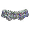
Abstract: Rhodobacter sphaeroides is a model organism in bacterial photosynthesis, and its light-harvesting-reaction center (LH1-RC) complex contains both dimeric and monomeric forms. Here we present cryo-EM ...Rhodobacter sphaeroides is a model organism in bacterial photosynthesis, and its light-harvesting-reaction center (LH1-RC) complex contains both dimeric and monomeric forms. Here we present cryo-EM structures of the native LH1-RC dimer and an LH1-RC monomer lacking protein-U (ΔU). The native dimer reveals several asymmetric features including the arrangement of its two monomeric components, the structural integrity of protein-U, the overall organization of LH1, and rigidities of the proteins and pigments. PufX plays a critical role in connecting the two monomers in a dimer, with one PufX interacting at its N-terminus with another PufX and an LH1 β-polypeptide in the other monomer. One protein-U was only partially resolved in the dimeric structure, signaling different degrees of disorder in the two monomers. The ΔU LH1-RC monomer was half-moon-shaped and contained 11 α- and 10 β-polypeptides, indicating a critical role for protein-U in controlling the number of αβ-subunits required for dimer assembly and stabilization. These features are discussed in relation to membrane topology and an assembly model proposed for the native dimeric complex.
History
DepositionNov 13, 2021-
Header (metadata) releaseApr 27, 2022-
Map releaseApr 27, 2022-
UpdateOct 30, 2024-
Current statusOct 30, 2024Processing site: PDBj / Status: Released

-
Structure visualization

Supplemental images

Downloads & links

-
Map

FileDownload / File: emd_32192.map.gz / Format: CCP4 / Size: 347.6 MB / Type: IMAGE STORED AS FLOATING POINT NUMBER (4 BYTES)
Projections & slices

Image control

Size
Brightness
Contrast
Others
AxesX (Sec.)Y (Row.)Z (Col.)
0.82 Å/pix.
x 450 pix.
= 369. Å
0.82 Å/pix.
x 450 pix.
= 369. Å
0.82 Å/pix.
x 450 pix.
= 369. Å

Surface

Projections

Slices (1/3)

Slices (1/2)

Slices (2/3)

Images are generated by Spider.

Voxel sizeX=Y=Z: 0.82 Å
Density
Contour LevelBy AUTHOR: 3.2
Minimum - Maximum-18.202724 - 39.517386999999999
Average (Standard dev.)0.0005062192 (±1.0001585)
SymmetrySpace group: 1
Details

EMDB XML:

Map geometry
Axis orderZYX
Origin000
Dimensions450450450
Spacing450450450
CellA=B=C: 369.0 Å
α=β=γ: 90.0 °

-
Supplemental data

-
Sample components

+
Entire : Photosynthetic LH1-RC complex from the purple phototrophic bacter...

EntireName: Photosynthetic LH1-RC complex from the purple phototrophic bacterium Rhodobacter sphaeroides dimer
Components
  • Complex: Photosynthetic LH1-RC complex from the purple phototrophic bacterium Rhodobacter sphaeroides dimer
    • Complex: Rhodobacter sphaeroides Dimeric LH1-RC Complex
      • Protein or peptide: Photosynthetic reaction center L subunit
      • Protein or peptide: Reaction center protein M chain
      • Protein or peptide: Photosynthetic reaction center subunit H
      • Protein or peptide: Antenna pigment protein alpha chain
      • Protein or peptide: Antenna pigment protein beta chain
      • Protein or peptide: PufX
      • Protein or peptide: protein-U
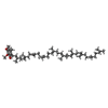
  • Ligand: BACTERIOCHLOROPHYLL A
  • Ligand: BACTERIOPHEOPHYTIN A
  • Ligand: UBIQUINONE-10
  • Ligand: DODECYL-BETA-D-MALTOSIDE
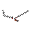
  • Ligand: (1R)-2-{[{[(2S)-2,3-DIHYDROXYPROPYL]OXY}(HYDROXY)PHOSPHORYL]OXY}-1-[(PALMITOYLOXY)METHYL]ETHYL (11E)-OCTADEC-11-ENOATE
  • Ligand: FE (III) ION
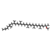
  • Ligand: SPHEROIDENE
  • Ligand: CARDIOLIPIN
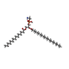
  • Ligand: PHOSPHATIDYLETHANOLAMINE
  • Ligand: LAURYL DIMETHYLAMINE-N-OXIDE

+
Supramolecule #1: Photosynthetic LH1-RC complex from the purple phototrophic bacter...

SupramoleculeName: Photosynthetic LH1-RC complex from the purple phototrophic bacterium Rhodobacter sphaeroides dimer
type: complex / ID: 1 / Parent: 0 / Macromolecule list: #1-#7
Source (natural)Organism: Rhodobacter sphaeroides f. sp. denitrificans (bacteria)
Strain: IL106

+
Supramolecule #2: Rhodobacter sphaeroides Dimeric LH1-RC Complex

SupramoleculeName: Rhodobacter sphaeroides Dimeric LH1-RC Complex / type: complex / ID: 2 / Parent: 1 / Macromolecule list: #1-#7
Source (natural)Organism: Rhodobacter sphaeroides f. sp. denitrificans (bacteria)
Strain: IL106

+
Macromolecule #1: Photosynthetic reaction center L subunit

MacromoleculeName: Photosynthetic reaction center L subunit / type: protein_or_peptide / ID: 1 / Number of copies: 2 / Enantiomer: LEVO
Source (natural)Organism: Rhodobacter sphaeroides f. sp. denitrificans (bacteria)
Molecular weightTheoretical: 31.360416 KDa
SequenceString: ALLSFERKYR VPGGTLVGGN LFDFWVGPFY VGFFGVATFF FAALGIILIA WSAVLQGTWN PQLISVYPPA LEYGLGGAPL AKGGLWQII TICATGAFVS WALREVEICR KLGIGYHIPF AFAFAILAYL TLVLFRPVMM GAWGYAFPYG IWTHLDWVSN T GYTYGNFH ...String:
ALLSFERKYR VPGGTLVGGN LFDFWVGPFY VGFFGVATFF FAALGIILIA WSAVLQGTWN PQLISVYPPA LEYGLGGAPL AKGGLWQII TICATGAFVS WALREVEICR KLGIGYHIPF AFAFAILAYL TLVLFRPVMM GAWGYAFPYG IWTHLDWVSN T GYTYGNFH YNPAHMIAIT FFFTNALALA LHGALVLSAA NPEKGKEMRT PDHEDTFFRD LVGYSIGTLG IHRLGLLLSL SA VFFSALC MIITGTIWFD QWVDWWQWWV KLPWWANIPG GING

UniProtKB: Reaction center protein L chain

+
Macromolecule #2: Reaction center protein M chain

MacromoleculeName: Reaction center protein M chain / type: protein_or_peptide / ID: 2 / Number of copies: 2 / Enantiomer: LEVO
Source (natural)Organism: Rhodobacter sphaeroides f. sp. denitrificans (bacteria)
Molecular weightTheoretical: 34.412566 KDa
SequenceString: AEYQNIFTQV QVRGPADLGM TEDVNLANRS GVGPFSTLLG WFGNAQLGPI YLGSLGVLSL FSGLMWFFTI GIWFWYQAGW NPAVFLRDL FFFSLEPPAP EYGLSFAAPL KEGGLWLIAS FFMFVAVWSW WGRTYLRAQA LGMGKHTAWA FLSAIWLWMV L GFIRPILM ...String:
AEYQNIFTQV QVRGPADLGM TEDVNLANRS GVGPFSTLLG WFGNAQLGPI YLGSLGVLSL FSGLMWFFTI GIWFWYQAGW NPAVFLRDL FFFSLEPPAP EYGLSFAAPL KEGGLWLIAS FFMFVAVWSW WGRTYLRAQA LGMGKHTAWA FLSAIWLWMV L GFIRPILM GSWSEAVPYG IFSHLDWTNN FSLVHGNLFY NPFHGLSIAF LYGSALLFAM HGATILAVSR FGGERELEQI AD RGTAAER AALFWRWTMG FNATMEGIHR WAIWMAVLVT LTGGIGILLS GTVVDNWYVW GQNHGMAPLN

UniProtKB: Reaction center protein M chain

+
Macromolecule #3: Photosynthetic reaction center subunit H

MacromoleculeName: Photosynthetic reaction center subunit H / type: protein_or_peptide / ID: 3 / Number of copies: 2 / Enantiomer: LEVO
Source (natural)Organism: Rhodobacter sphaeroides f. sp. denitrificans (bacteria)
Molecular weightTheoretical: 28.09135 KDa
SequenceString: MVGVTAFGNF DLASLAIYSF WIFLAGLIYY LQTENMREGY PLENEDGTPA ANQGPFPLPK PKTFILPHGR GTLTVPGPES EDRPIALAR TAVSEGFPHA PTGDPMKDGV GPASWVARRD LPELDGHGHN KIKPMKAAAG FYVSAGKNPI GLPVRGCDLE I AGKVVDIW ...String:
MVGVTAFGNF DLASLAIYSF WIFLAGLIYY LQTENMREGY PLENEDGTPA ANQGPFPLPK PKTFILPHGR GTLTVPGPES EDRPIALAR TAVSEGFPHA PTGDPMKDGV GPASWVARRD LPELDGHGHN KIKPMKAAAG FYVSAGKNPI GLPVRGCDLE I AGKVVDIW VDIPEQMARF LEVELKDGST RLLPMQMVKV QSNRVHVNAL SSDLFAGIPT IKSPTEVTLL EEDKICGYVA GG LMYAAPK RKSVVAAMLA EYA

UniProtKB: Photosynthetic reaction center subunit H

+
Macromolecule #4: Antenna pigment protein alpha chain

MacromoleculeName: Antenna pigment protein alpha chain / type: protein_or_peptide / ID: 4 / Number of copies: 28 / Enantiomer: LEVO
Source (natural)Organism: Rhodobacter sphaeroides f. sp. denitrificans (bacteria)
Molecular weightTheoretical: 6.47378 KDa
SequenceString:
(FME)SKFYKIWMI FDPRRVFVAQ GVFLFLLAVM IHLILLSTPS YNWLEISAAK YNRV

UniProtKB: Antenna pigment protein alpha chain

+
Macromolecule #5: Antenna pigment protein beta chain

MacromoleculeName: Antenna pigment protein beta chain / type: protein_or_peptide / ID: 5 / Number of copies: 28 / Enantiomer: LEVO
Source (natural)Organism: Rhodobacter sphaeroides f. sp. denitrificans (bacteria)
Molecular weightTheoretical: 5.461166 KDa
SequenceString:
ADKSDLGYTG LTDEQAQELH SVYMSGLWLF SAVAIVAHLA VYIWRPWF

UniProtKB: Antenna pigment protein beta chain

+
Macromolecule #6: PufX

MacromoleculeName: PufX / type: protein_or_peptide / ID: 6 / Number of copies: 2 / Enantiomer: LEVO
Source (natural)Organism: Rhodobacter sphaeroides f. sp. denitrificans (bacteria)
Molecular weightTheoretical: 8.886395 KDa
SequenceString:
ADKTIFNDHL NTNPKTNLRL WVAFQMMKGA GWAGGVFFGT LLLIGFFRVV GRMLPIDENP APAPNITGAL ETGIELIKHL V

UniProtKB: Intrinsic membrane protein PufX

+
Macromolecule #7: protein-U

MacromoleculeName: protein-U / type: protein_or_peptide / ID: 7 / Number of copies: 2 / Enantiomer: LEVO
Source (natural)Organism: Rhodobacter sphaeroides f. sp. denitrificans (bacteria)
Molecular weightTheoretical: 5.555558 KDa
SequenceString:
MPEVSEFAFR LMMAAVIFVG VGIMFAFAGG HWFVGLVVGG LVAAFFAATP NSN

UniProtKB: Uncharacterized protein

+
Macromolecule #8: BACTERIOCHLOROPHYLL A

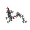
MacromoleculeName: BACTERIOCHLOROPHYLL A / type: ligand / ID: 8 / Number of copies: 64 / Formula: BCL
Molecular weightTheoretical: 911.504 Da
Chemical component information

ChemComp-BCL:
BACTERIOCHLOROPHYLL A

+
Macromolecule #9: BACTERIOPHEOPHYTIN A

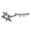
MacromoleculeName: BACTERIOPHEOPHYTIN A / type: ligand / ID: 9 / Number of copies: 4 / Formula: BPH
Molecular weightTheoretical: 889.215 Da
Chemical component information

ChemComp-BPH:
BACTERIOPHEOPHYTIN A

+
Macromolecule #10: UBIQUINONE-10

MacromoleculeName: UBIQUINONE-10 / type: ligand / ID: 10 / Number of copies: 8 / Formula: U10
Molecular weightTheoretical: 863.343 Da
Chemical component information

ChemComp-U10:
UBIQUINONE-10

+
Macromolecule #11: DODECYL-BETA-D-MALTOSIDE

MacromoleculeName: DODECYL-BETA-D-MALTOSIDE / type: ligand / ID: 11 / Number of copies: 27 / Formula: LMT
Molecular weightTheoretical: 510.615 Da
Chemical component information

ChemComp-LMT:
DODECYL-BETA-D-MALTOSIDE / detergent*YM

+
Macromolecule #12: (1R)-2-{[{[(2S)-2,3-DIHYDROXYPROPYL]OXY}(HYDROXY)PHOSPHORYL]OXY}-...

MacromoleculeName: (1R)-2-{[{[(2S)-2,3-DIHYDROXYPROPYL]OXY}(HYDROXY)PHOSPHORYL]OXY}-1-[(PALMITOYLOXY)METHYL]ETHYL (11E)-OCTADEC-11-ENOATE
type: ligand / ID: 12 / Number of copies: 26 / Formula: PGV
Molecular weightTheoretical: 749.007 Da
Chemical component information

ChemComp-PGV:
(1R)-2-{[{[(2S)-2,3-DIHYDROXYPROPYL]OXY}(HYDROXY)PHOSPHORYL]OXY}-1-[(PALMITOYLOXY)METHYL]ETHYL (11E)-OCTADEC-11-ENOATE / phospholipid*YM

+
Macromolecule #13: FE (III) ION

MacromoleculeName: FE (III) ION / type: ligand / ID: 13 / Number of copies: 2 / Formula: FE
Molecular weightTheoretical: 55.845 Da

+
Macromolecule #14: SPHEROIDENE

MacromoleculeName: SPHEROIDENE / type: ligand / ID: 14 / Number of copies: 54 / Formula: SPO
Molecular weightTheoretical: 568.914 Da
Chemical component information

ChemComp-7OT:
SPHEROIDENE

+
Macromolecule #15: CARDIOLIPIN

MacromoleculeName: CARDIOLIPIN / type: ligand / ID: 15 / Number of copies: 11 / Formula: CDL
Molecular weightTheoretical: 1.464043 KDa
Chemical component information

ChemComp-CDL:
CARDIOLIPIN / phospholipid*YM

+
Macromolecule #16: PHOSPHATIDYLETHANOLAMINE

MacromoleculeName: PHOSPHATIDYLETHANOLAMINE / type: ligand / ID: 16 / Number of copies: 4 / Formula: PTY
Molecular weightTheoretical: 734.039 Da
Chemical component information

ChemComp-PTY:
PHOSPHATIDYLETHANOLAMINE / phospholipid*YM

+
Macromolecule #17: LAURYL DIMETHYLAMINE-N-OXIDE

MacromoleculeName: LAURYL DIMETHYLAMINE-N-OXIDE / type: ligand / ID: 17 / Number of copies: 1 / Formula: LDA
Molecular weightTheoretical: 229.402 Da
Chemical component information

ChemComp-LDA:
LAURYL DIMETHYLAMINE-N-OXIDE / LDAO, detergent*YM

-
Experimental details

-
Structure determination

Methodcryo EM
Processingsingle particle reconstruction
Aggregation stateparticle

-
Sample preparation

Concentration3.0 mg/mL
BufferpH: 8
VitrificationCryogen name: ETHANE / Chamber humidity: 80 % / Chamber temperature: 277 K / Instrument: LEICA EM GP
DetailsThis sample was monodisperse.

-
Electron microscopy

MicroscopeFEI TITAN KRIOS
Image recordingFilm or detector model: FEI FALCON III (4k x 4k) / Detector mode: COUNTING / Average exposure time: 1.275 sec. / Average electron dose: 42.0 e/Å2
Electron beamAcceleration voltage: 300 kV / Electron source: FIELD EMISSION GUN
Electron opticsIllumination mode: FLOOD BEAM / Imaging mode: BRIGHT FIELD / Nominal defocus max: 3.0 µm / Nominal defocus min: 1.0 µm
Sample stageSpecimen holder model: FEI TITAN KRIOS AUTOGRID HOLDER / Cooling holder cryogen: NITROGEN
Experimental equipment
Model: Titan Krios / Image courtesy: FEI Company

+
Image processing

Particle selectionNumber selected: 164496
Startup modelType of model: NONE
Final reconstructionApplied symmetry - Point group: C1 (asymmetric) / Algorithm: FOURIER SPACE / Resolution.type: BY AUTHOR / Resolution: 2.75 Å / Resolution method: FSC 0.143 CUT-OFF / Software - Name: RELION (ver. 3.1) / Number images used: 124916
Initial angle assignmentType: NOT APPLICABLE
Final angle assignmentType: MAXIMUM LIKELIHOOD / Software - Name: RELION (ver. 3.1)
Final 3D classificationSoftware - Name: RELION (ver. 3.1)
FSC plot (resolution estimation)

-
Atomic model buiding 1

Initial modelPDB ID:

Chain - Source name: PDB / Chain - Initial model type: experimental model
RefinementSpace: REAL / Protocol: RIGID BODY FIT / Overall B value: 60 / Target criteria: Correlation coefficient
Output model

PDB-7vy2:
STRUCTURE OF PHOTOSYNTHETIC LH1-RC SUPER-COMPLEX OF RHODOBACTER SPHAEROIDES DIMER

+
About Yorodumi

-
News

-
Feb 9, 2022. New format data for meta-information of EMDB entries

New format data for meta-information of EMDB entries

  • Version 3 of the EMDB header file is now the official format.
  • The previous official version 1.9 will be removed from the archive.

Related info.:EMDB header

External links:wwPDB to switch to version 3 of the EMDB data model

-
Aug 12, 2020. Covid-19 info

Covid-19 info

URL: https://pdbj.org/emnavi/covid19.php

New page: Covid-19 featured information page in EM Navigator.

Related info.:Covid-19 info / Mar 5, 2020. Novel coronavirus structure data

+
Mar 5, 2020. Novel coronavirus structure data

Novel coronavirus structure data

Related info.:Yorodumi Speices / Aug 12, 2020. Covid-19 info

External links:COVID-19 featured content - PDBj / Molecule of the Month (242):Coronavirus Proteases

+
Jan 31, 2019. EMDB accession codes are about to change! (news from PDBe EMDB page)

EMDB accession codes are about to change! (news from PDBe EMDB page)

  • The allocation of 4 digits for EMDB accession codes will soon come to an end. Whilst these codes will remain in use, new EMDB accession codes will include an additional digit and will expand incrementally as the available range of codes is exhausted. The current 4-digit format prefixed with “EMD-” (i.e. EMD-XXXX) will advance to a 5-digit format (i.e. EMD-XXXXX), and so on. It is currently estimated that the 4-digit codes will be depleted around Spring 2019, at which point the 5-digit format will come into force.
  • The EM Navigator/Yorodumi systems omit the EMD- prefix.

Related info.:Q: What is EMD? / ID/Accession-code notation in Yorodumi/EM Navigator

External links:EMDB Accession Codes are Changing Soon! / Contact to PDBj

+
Jul 12, 2017. Major update of PDB

Major update of PDB

  • wwPDB released updated PDB data conforming to the new PDBx/mmCIF dictionary.
  • This is a major update changing the version number from 4 to 5, and with Remediation, in which all the entries are updated.
  • In this update, many items about electron microscopy experimental information are reorganized (e.g. em_software).
  • Now, EM Navigator and Yorodumi are based on the updated data.

External links:wwPDB Remediation / Enriched Model Files Conforming to OneDep Data Standards Now Available in the PDB FTP Archive

-
Yorodumi

Thousand views of thousand structures

  • Yorodumi is a browser for structure data from EMDB, PDB, SASBDB, etc.
  • This page is also the successor to EM Navigator detail page, and also detail information page/front-end page for Omokage search.
  • The word "yorodu" (or yorozu) is an old Japanese word meaning "ten thousand". "mi" (miru) is to see.

Related info.:EMDB / PDB / SASBDB / Comparison of 3 databanks / Yorodumi Search / Aug 31, 2016. New EM Navigator & Yorodumi / Yorodumi Papers / Jmol/JSmol / Function and homology information / Changes in new EM Navigator and Yorodumi

Read more