 ムービー
ムービー コントローラー


+ データを開く

- 基本情報
基本情報
| 登録情報 | データベース: EMDB / ID: EMD-3193 | |||||||||
|---|---|---|---|---|---|---|---|---|---|---|
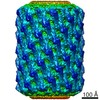
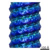
| タイトル | Helical reconstruction of amphiphysin N-BAR with a membrane tube radius of 156 Angstrom by cryo-electron microscopy | |||||||||
 マップデータ マップデータ | Helical reconstruction of amphiphysin N-BAR with a membrane tube diameter of 156 Angstrom. | |||||||||
 試料 試料 |
| |||||||||
 キーワード キーワード | amphiphysin / N-BAR / amphiphysin/BIN1 / BIN1 | |||||||||
| 機能・相同性 |  機能・相同性情報 機能・相同性情報rhabdomere membrane biogenesis / organelle / rhabdomere development / Clathrin-mediated endocytosis / Neutrophil degranulation / regulation of muscle contraction / exocytosis / cleavage furrow / phospholipid binding / protein localization ...rhabdomere membrane biogenesis / organelle / rhabdomere development / Clathrin-mediated endocytosis / Neutrophil degranulation / regulation of muscle contraction / exocytosis / cleavage furrow / phospholipid binding / protein localization / synapse / plasma membrane / cytoplasm 類似検索 - 分子機能 | |||||||||
| 生物種 |  | |||||||||
| 手法 | らせん対称体再構成法 / クライオ電子顕微鏡法 / 解像度: 11.2 Å | |||||||||
 データ登録者 データ登録者 | Adam J / Basnet N / Mizuno N | |||||||||
 引用 引用 | ジャーナル: Sci Rep / 年: 2015 タイトル: Structural insights into the cooperative remodeling of membranes by amphiphysin/BIN1. 著者: Julia Adam / Nirakar Basnet / Naoko Mizuno /  要旨: Amphiphysin2/BIN1 is a crescent-shaped N-BAR protein playing a key role in forming deeply invaginated tubes in muscle T-tubules. Amphiphysin2/BIN1 structurally stabilizes tubular formations in ...Amphiphysin2/BIN1 is a crescent-shaped N-BAR protein playing a key role in forming deeply invaginated tubes in muscle T-tubules. Amphiphysin2/BIN1 structurally stabilizes tubular formations in contrast to other N-BAR proteins involved in dynamic membrane scission processes; however, the molecular mechanism of the stabilizing effect is poorly understood. Using cryo-EM, we investigated the assembly of the amphiphysin/BIN1 on a membrane tube. We found that the N-BAR domains self-assemble on the membrane surface in a highly cooperative manner. Our biochemical assays and 3D reconstructions indicate that the N-terminal amphipathic helix H0 plays an important role in the initiation of the tube assembly and further in organizing BAR-mediated polymerization by locking adjacent N-BAR domains. Mutants that lack H0 or the tip portion, which is also involved in interactions of the neighboring BAR unit, lead to a disruption of the polymer organization, even though tubulation can still be observed. The regulatory region of amphiphysin/BIN1 including an SH3 domain does not have any apparent involvement in the polymer lattice. Our study indicates that the H0 helix and the BAR tip are necessary for efficient and organized self-assembly of amphiphysin/N-BAR. | |||||||||
| 履歴 |
|
- 構造の表示
構造の表示
| ムービー |
ムービービューア |
|---|---|
| 構造ビューア | EMマップ: SurfViewMolmilJmol/JSmol |
| 添付画像 |

- ダウンロードとリンク
-EMDBアーカイブ
| マップデータ | emd_3193.map.gz | 90.3 MB | EMDBマップデータ形式 | |
|---|---|---|---|---|
| ヘッダ (付随情報) | emd-3193-v30.xmlemd-3193.xml | 10.5 KB 10.5 KB | 表示 表示 | EMDBヘッダ |
| 画像 | 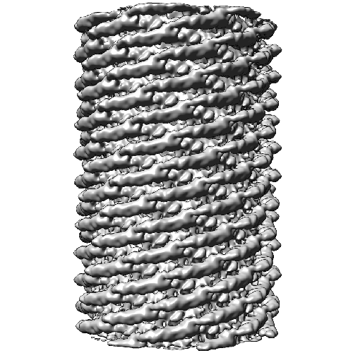 3193emdb13509_500x500.png 3193emdb13509_500x500.png | 978.8 KB | ||
| アーカイブディレクトリ |  http://ftp.pdbj.org/pub/emdb/structures/EMD-3193ftp://ftp.pdbj.org/pub/emdb/structures/EMD-3193 http://ftp.pdbj.org/pub/emdb/structures/EMD-3193ftp://ftp.pdbj.org/pub/emdb/structures/EMD-3193 | HTTPS FTP |
-検証レポート
| 文書・要旨 | emd_3193_validation.pdf.gz | 270.1 KB | 表示 | EMDB検証レポート |
|---|---|---|---|---|
| 文書・詳細版 | emd_3193_full_validation.pdf.gz | 269.2 KB | 表示 | |
| XML形式データ | emd_3193_validation.xml.gz | 6 KB | 表示 | |
| アーカイブディレクトリ | https://ftp.pdbj.org/pub/emdb/validation_reports/EMD-3193ftp://ftp.pdbj.org/pub/emdb/validation_reports/EMD-3193 | HTTPS FTP |
-関連構造データ


-リンク
| EMDBのページ | EMDB (EBI/PDBe) / EMDataResource |
|---|---|
| 「今月の分子」の関連する項目 |


-マップ
| ファイル | ダウンロード / ファイル: emd_3193.map.gz / 形式: CCP4 / 大きさ: 100.6 MB / タイプ: IMAGE STORED AS FLOATING POINT NUMBER (4 BYTES) | ||||||||||||||||||||||||||||||||||||||||||||||||||||||||||||
|---|---|---|---|---|---|---|---|---|---|---|---|---|---|---|---|---|---|---|---|---|---|---|---|---|---|---|---|---|---|---|---|---|---|---|---|---|---|---|---|---|---|---|---|---|---|---|---|---|---|---|---|---|---|---|---|---|---|---|---|---|---|
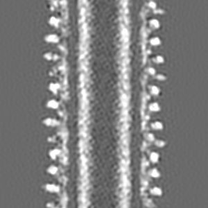
| 注釈 | Helical reconstruction of amphiphysin N-BAR with a membrane tube diameter of 156 Angstrom. | ||||||||||||||||||||||||||||||||||||||||||||||||||||||||||||
| 投影像・断面図 | 画像のコントロール
画像は Spider により作成 | ||||||||||||||||||||||||||||||||||||||||||||||||||||||||||||
| ボクセルのサイズ | X=Y=Z: 1.82 Å | ||||||||||||||||||||||||||||||||||||||||||||||||||||||||||||
| 密度 |
| ||||||||||||||||||||||||||||||||||||||||||||||||||||||||||||
| 対称性 | 空間群: 1 | ||||||||||||||||||||||||||||||||||||||||||||||||||||||||||||
| 詳細 | EMDB XML:
CCP4マップ ヘッダ情報:
| ||||||||||||||||||||||||||||||||||||||||||||||||||||||||||||
 Z (Sec.)
 Y (Row.)
 X (Col.)


-添付データ
- 試料の構成要素
-全体 : Amphiphysin N-BAR with a membrane tube radius of 156 Angstrom
| 全体 | 名称: Amphiphysin N-BAR with a membrane tube radius of 156 Angstrom |
|---|---|
| 要素 |
|
-超分子 #1000: Amphiphysin N-BAR with a membrane tube radius of 156 Angstrom
| 超分子 | 名称: Amphiphysin N-BAR with a membrane tube radius of 156 Angstrom タイプ: sample / ID: 1000 / 集合状態: polymer / Number unique components: 1 |
|---|
-分子 #1: Amphiphysin N-BAR
| 分子 | 名称: Amphiphysin N-BAR / タイプ: protein_or_peptide / ID: 1 / Name.synonym: Amph N-BAR / 集合状態: Dimer / 組換発現: Yes |
|---|---|
| 由来(天然) | 生物種: 別称: Fruit Fly / 細胞中の位置: T-Tubules |
| 組換発現 | 生物種:  |
| 配列 | UniProtKB: Amphiphysin / InterPro: Amphiphysin |
-実験情報
-構造解析
| 手法 | クライオ電子顕微鏡法 |
|---|---|
 解析 解析 | らせん対称体再構成法 |
| 試料の集合状態 | helical array |
-試料調製
| 濃度 | 0.6 mg/mL |
|---|---|
| 緩衝液 | pH: 7.4 / 詳細: 20 mM Hepes, 500 mM NaCl, 1 mM EDTA, 1 mM DTT |
| グリッド | 詳細: Quantifoil R 2/1, Cu, 300 mesh grids |
| 凍結 | 凍結剤: ETHANE / 装置: HOMEMADE PLUNGER / 手法: 10 s of manual blotting before plunging |
| 詳細 | 20 uM of Amph N-BAR protein was incubated with 720 uM liposomes at RT. |
- 電子顕微鏡法
| 顕微鏡 | FEI POLARA 300 |
|---|---|
| 特殊光学系 | エネルギーフィルター - 名称: GIF 200 |
| 日付 | 2014年8月19日 |
| 撮影 | カテゴリ: CCD / フィルム・検出器のモデル: GATAN K2 (4k x 4k) / 実像数: 271 / 平均電子線量: 30 e/Å2 詳細: every image is average of 30 frames recorded by the direct electron detector |
| 電子線 | 加速電圧: 300 kV / 電子線源:  FIELD EMISSION GUN FIELD EMISSION GUN |
| 電子光学系 | 照射モード: FLOOD BEAM / 撮影モード: BRIGHT FIELD / Cs: 2.0 mm / 最大 デフォーカス(公称値): 3.2 µm / 最小 デフォーカス(公称値): 0.5 µm / 倍率(公称値): 62000 |
| 試料ステージ | 試料ホルダーモデル: OTHER |
| 実験機器 |  モデル: Tecnai Polara / 画像提供: FEI Company |
-画像解析
| 詳細 | Helix handedness is not confirmed by sub-tomogram averaging. Used programs: BSOFT software package, particle picking by EMAN2 with e2helixboxer, 2D classification by Relion, 3D helical reconstruction by IHRSR implemented into SPIDER. |
|---|---|
| 最終 再構成 | 想定した対称性 - らせんパラメータ - Δz: 3.83 Å 想定した対称性 - らせんパラメータ - ΔΦ: 55.96 ° 想定した対称性 - らせんパラメータ - 軸対称性: C1 (非対称) アルゴリズム: OTHER / 解像度のタイプ: BY AUTHOR / 解像度: 11.2 Å / 解像度の算出法: OTHER ソフトウェア - 名称: BSOFT, EMAN2, Relion, SPIDER, IHRSR 詳細: For the reconstruction 1392 segmented particles were used. The particles were 2D classified by Relion and for the reconstruction the helical symmetry was applied using IHRSR. |
| CTF補正 | 詳細: Phases of individual images are flipped |
-原子モデル構築 1
| 初期モデル | PDB ID: |
|---|---|
| ソフトウェア | 名称: Chimera |
| 詳細 | The amphiphysin N-BAR dimer was fitted by manual docking using Chimera. |
| 精密化 | 空間: REAL / プロトコル: RIGID BODY FIT |

