- EMDB-30547: Cryo-EM Structure of PSII at 1.95 angstrom resolution -
+
データを開く
IDまたはキーワード:
読み込み中...
-
基本情報
登録情報
データベース: EMDB / ID: EMD-30547
タイトル
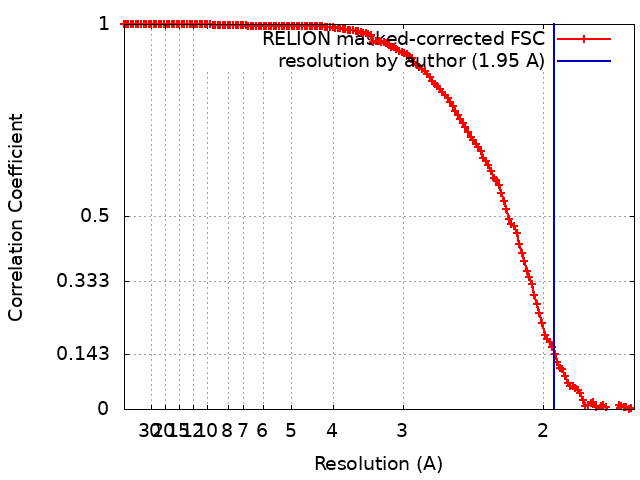
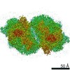
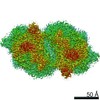
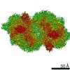
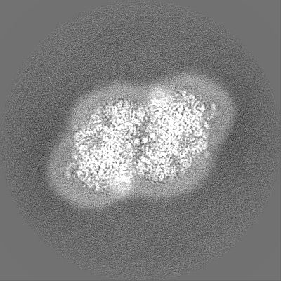
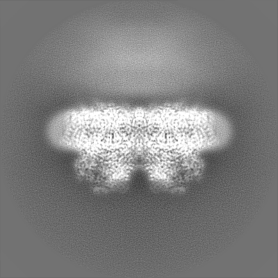
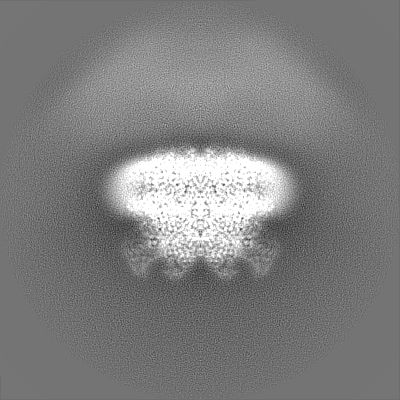
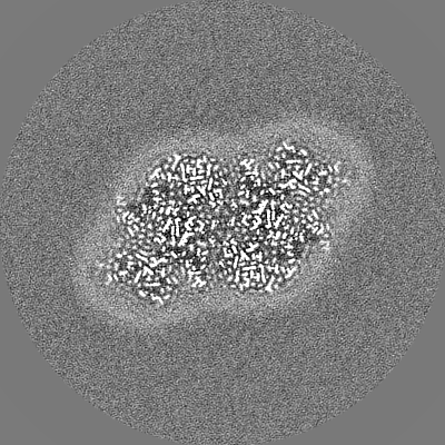
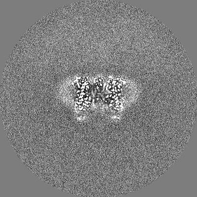
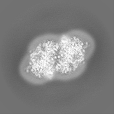
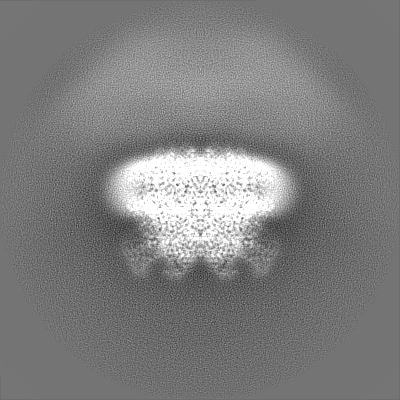
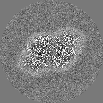
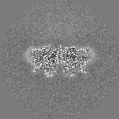
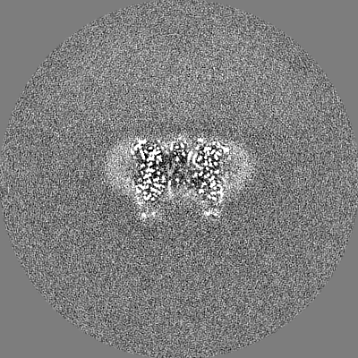
Cryo-EM Structure of PSII at 1.95 angstrom resolution
マップデータ
試料
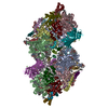
複合体: PSII dimer
タンパク質・ペプチド: x 20種
リガンド: x 19種
キーワード
Photosystem II / PHOTOSYNTHESIS
機能・相同性
機能・相同性情報
photosystem II oxygen evolving complex / photosystem II assembly / oxygen evolving activity / photosystem II stabilization / photosystem II reaction center / photosystem II / oxidoreductase activity, acting on diphenols and related substances as donors, oxygen as acceptor / photosynthetic electron transport chain / photosystem II / response to herbicide ...photosystem II oxygen evolving complex / photosystem II assembly / oxygen evolving activity / photosystem II stabilization / photosystem II reaction center / photosystem II / oxidoreductase activity, acting on diphenols and related substances as donors, oxygen as acceptor / photosynthetic electron transport chain / photosystem II / response to herbicide / extrinsic component of membrane / chlorophyll binding / plasma membrane-derived thylakoid membrane / photosynthetic electron transport in photosystem II / photosynthesis, light reaction / phosphate ion binding / : / photosynthesis / respiratory electron transport chain / manganese ion binding / electron transfer activity / protein stabilization / iron ion binding / heme binding / metal ion binding 類似検索 - 分子機能
Photosystem II protein Y (PsbY) / Photosystem II PsbY / Photosystem II PsbU, oxygen evolving complex / Photosystem II 12 kDa extrinsic protein (PsbU) / Photosystem II PsbV, cytochrome c-550 precursor / Photosystem II cytochrome c-550 precursor / Cytochrome c-550 domain / Cytochrome c-550 domain / Photosystem II PsbJ / Photosystem II PsbJ superfamily ...Photosystem II protein Y (PsbY) / Photosystem II PsbY / Photosystem II PsbU, oxygen evolving complex / Photosystem II 12 kDa extrinsic protein (PsbU) / Photosystem II PsbV, cytochrome c-550 precursor / Photosystem II cytochrome c-550 precursor / Cytochrome c-550 domain / Cytochrome c-550 domain / Photosystem II PsbJ / Photosystem II PsbJ superfamily / PsbJ / Photosystem II PsbX, type 1 subfamily / Photosystem II PsbO, manganese-stabilising / Manganese-stabilising protein / photosystem II polypeptide / Photosystem II reaction centre protein Ycf12 / Photosystem II complex subunit Ycf12 / Photosystem II reaction centre M protein (PsbM) / Photosystem II PsbM superfamily / Photosystem II PsbM / Photosystem II PsbZ, reaction centre / Photosystem II PsbZ superfamily / YCF9 / Photosystem II PsbX / Photosystem II reaction centre X protein (PsbX) / Photosystem II PsbT / Photosystem II PsbL / Photosystem II PsbL superfamily / Photosystem II PsbT superfamily / Photosystem II reaction centre T protein / PsbL protein / Photosystem II PsbK / Photosystem II CP43 reaction centre protein / Photosystem II PsbK superfamily / Photosystem II CP43 reaction centre protein superfamily / Photosystem II 4 kDa reaction centre component / Photosystem II PsbI / Photosystem II CP47 reaction centre protein / Photosystem II PsbI superfamily / Photosystem II reaction centre I protein (PSII 4.8 kDa protein) / Photosystem II reaction centre protein H / Photosystem II reaction centre protein H superfamily / Photosystem II 10 kDa phosphoprotein / Photosystem II protein D1 / Photosystem II D2 protein / Photosystem II cytochrome b559, conserved site / Photosystem II cytochrome b559, alpha subunit / Photosystem II cytochrome b559, beta subunit / Photosystem II cytochrome b559, N-terminal / Photosystem II cytochrome b559, alpha subunit, lumenal region / Photosystem II cytochrome b559, alpha subunit superfamily / Cytochrome b559, alpha (gene psbE) and beta (gene psbF)subunits / Lumenal portion of Cytochrome b559, alpha (gene psbE) subunit / Cytochrome b559 subunits heme-binding site signature. / : / Photosystem antenna protein-like / Photosystem antenna protein-like superfamily / Photosystem II protein / Outer membrane protein/outer membrane enzyme PagP, beta-barrel / : / Photosynthetic reaction centre, L/M / Photosystem II protein D1/D2 superfamily / Photosynthetic reaction centre protein / Photosynthetic reaction center proteins signature. / Cytochrome c family profile. / Cytochrome c-like domain / Cytochrome c-like domain superfamily 類似検索 - ドメイン・相同性
Photosystem II CP47 reaction center protein / Photosystem II extrinsic protein O / Photosystem II reaction center protein Psb30 / Photosystem II reaction center protein X / Photosystem II reaction center protein Z / Photosystem II CP43 reaction center protein / Photosystem II D2 protein / Photosystem II extrinsic protein V / Photosystem II reaction center protein Y / Cytochrome b559 subunit alpha ...Photosystem II CP47 reaction center protein / Photosystem II extrinsic protein O / Photosystem II reaction center protein Psb30 / Photosystem II reaction center protein X / Photosystem II reaction center protein Z / Photosystem II CP43 reaction center protein / Photosystem II D2 protein / Photosystem II extrinsic protein V / Photosystem II reaction center protein Y / Cytochrome b559 subunit alpha / Cytochrome b559 subunit beta / Photosystem II reaction center protein I / Photosystem II reaction center protein L / Photosystem II reaction center protein M / Photosystem II reaction center protein T / Photosystem II reaction center protein H / Photosystem II reaction center protein K / Photosystem II protein D1 / Photosystem II extrinsic protein U / Photosystem II reaction center protein J 類似検索 - 構成要素
ジャーナル: Commun Biol / 年: 2021 タイトル: High-resolution cryo-EM structure of photosystem II reveals damage from high-dose electron beams. 著者: Koji Kato / Naoyuki Miyazaki / Tasuku Hamaguchi / Yoshiki Nakajima / Fusamichi Akita / Koji Yonekura / Jian-Ren Shen / 要旨: Photosystem II (PSII) plays a key role in water-splitting and oxygen evolution. X-ray crystallography has revealed its atomic structure and some intermediate structures. However, these structures are ...Photosystem II (PSII) plays a key role in water-splitting and oxygen evolution. X-ray crystallography has revealed its atomic structure and some intermediate structures. However, these structures are in the crystalline state and its final state structure has not been solved. Here we analyzed the structure of PSII in solution at 1.95 Å resolution by single-particle cryo-electron microscopy (cryo-EM). The structure obtained is similar to the crystal structure, but a PsbY subunit was visible in the cryo-EM structure, indicating that it represents its physiological state more closely. Electron beam damage was observed at a high-dose in the regions that were easily affected by redox states, and reducing the beam dosage by reducing frames from 50 to 2 yielded a similar resolution but reduced the damage remarkably. This study will serve as a good indicator for determining damage-free cryo-EM structures of not only PSII but also all biological samples, especially redox-active metalloproteins.
EMPIAR-10556 (タイトル: Cryo-EM Structure of PSII at 1.95 angstrom resolution Data size: 427.6 Data #1: Unaligned multi-frame micrographs of PSII recorded by CRYO ARM 300 [micrographs - multiframe])
 ムービー
ムービー コントローラー


 データを開く
 基本情報
基本情報 マップデータ
 試料
試料 キーワード
キーワード 機能・相同性情報
 Thermosynechococcus vulcanus (バクテリア)
 データ登録者
 引用
引用
 構造の表示
構造の表示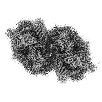
 ダウンロードとリンク
 emd_30547.png
 http://ftp.pdbj.org/pub/emdb/structures/EMD-30547


 Z (Sec.)
 Y (Row.)
 X (Col.)


 試料の構成要素


 解析
解析 電子顕微鏡法
 FIELD EMISSION GUN