 Movie
Movie Controller


+ Open data

- Basic information
| Entry | Database: EMDB / ID: EMD-30038 | |||||||||
|---|---|---|---|---|---|---|---|---|---|---|
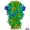
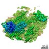
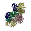
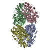
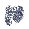
| Title | Cryo-EM structures of SADS-CoV spike glycoproteins | |||||||||
 Map data Map data | ||||||||||
 Sample Sample |
| |||||||||
 Keywords Keywords | VIRAL PROTEIN | |||||||||
| Function / homology |  Function and homology information Function and homology informationreceptor-mediated virion attachment to host cell / endocytosis involved in viral entry into host cell / fusion of virus membrane with host plasma membrane / fusion of virus membrane with host endosome membrane / viral envelope / virion membrane / membrane Similarity search - Function | |||||||||
| Biological species |  Swine acute diarrhea syndrome coronavirus Swine acute diarrhea syndrome coronavirus | |||||||||
| Method | single particle reconstruction / cryo EM / Resolution: 2.83 Å | |||||||||
 Authors Authors | Wang X / Yu J / Qiao S / Guo R | |||||||||
 Citation Citation | Journal: Nat Commun / Year: 2020 Title: Cryo-EM structures of HKU2 and SADS-CoV spike glycoproteins provide insights into coronavirus evolution. Authors: Jinfang Yu / Shuyuan Qiao / Runyu Guo / Xinquan Wang /  Abstract: Porcine coronavirus SADS-CoV has been identified from suckling piglets with severe diarrhea in southern China in 2017. The SADS-CoV genome shares ~95% identity to that of bat α-coronavirus HKU2, ...Porcine coronavirus SADS-CoV has been identified from suckling piglets with severe diarrhea in southern China in 2017. The SADS-CoV genome shares ~95% identity to that of bat α-coronavirus HKU2, suggesting that SADS-CoV may have emerged from a natural reservoir in bats. Here we report the cryo-EM structures of HKU2 and SADS-CoV spike (S) glycoprotein trimers at 2.38 Å and 2.83 Å resolution, respectively. We systematically compare the domains of HKU2 spike with those of α-, β-, γ-, and δ-coronavirus spikes, showing that the S1 subunit N- and C-terminal domains of HKU2/SADS-CoV are ancestral domains in the evolution of coronavirus spike proteins. The connecting region after the fusion peptide in the S2 subunit of HKU2/SADS-CoV adopts a unique conformation. These results structurally demonstrate a close evolutionary relationship between HKU2/SADS-CoV and β-coronavirus spikes and provide insights into the evolution and cross-species transmission of coronaviruses. | |||||||||
| History |
|
- Structure visualization
| Movie |
Movie viewer |
|---|---|
| Structure viewer | EM map: SurfViewMolmilJmol/JSmol |
| Supplemental images |

- Downloads & links
-EMDB archive
| Map data | emd_30038.map.gz | 7.2 MB | EMDB map data format | |
|---|---|---|---|---|
| Header (meta data) | emd-30038-v30.xmlemd-30038.xml | 9.7 KB 9.7 KB | Display Display | EMDB header |
| Images | 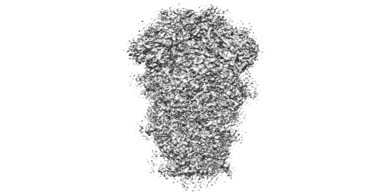 emd_30038.png emd_30038.png | 226.7 KB | ||
| Filedesc metadata | emd-30038.cif.gz | 5.6 KB | ||
| Archive directory |  http://ftp.pdbj.org/pub/emdb/structures/EMD-30038ftp://ftp.pdbj.org/pub/emdb/structures/EMD-30038 http://ftp.pdbj.org/pub/emdb/structures/EMD-30038ftp://ftp.pdbj.org/pub/emdb/structures/EMD-30038 | HTTPS FTP |
-Related structure data
| Related structure data |  6m16MC  6m15C M: atomic model generated by this map C: citing same article ( |
|---|---|
| Similar structure data |


-Links
| EMDB pages | EMDB (EBI/PDBe) / EMDataResource |
|---|
-Map
| File | Download / File: emd_30038.map.gz / Format: CCP4 / Size: 64 MB / Type: IMAGE STORED AS FLOATING POINT NUMBER (4 BYTES) | ||||||||||||||||||||||||||||||||||||||||||||||||||||||||||||||||||||
|---|---|---|---|---|---|---|---|---|---|---|---|---|---|---|---|---|---|---|---|---|---|---|---|---|---|---|---|---|---|---|---|---|---|---|---|---|---|---|---|---|---|---|---|---|---|---|---|---|---|---|---|---|---|---|---|---|---|---|---|---|---|---|---|---|---|---|---|---|---|
| Projections & slices | Image control
Images are generated by Spider. | ||||||||||||||||||||||||||||||||||||||||||||||||||||||||||||||||||||
| Voxel size | X=Y=Z: 1.061 Å | ||||||||||||||||||||||||||||||||||||||||||||||||||||||||||||||||||||
| Density |
| ||||||||||||||||||||||||||||||||||||||||||||||||||||||||||||||||||||
| Symmetry | Space group: 1 | ||||||||||||||||||||||||||||||||||||||||||||||||||||||||||||||||||||
| Details | EMDB XML:
CCP4 map header:
| ||||||||||||||||||||||||||||||||||||||||||||||||||||||||||||||||||||
 Z (Sec.)
 Y (Row.)
 X (Col.)


-Supplemental data
- Sample components
-Entire : SADS-CoV glycoprotein
| Entire | Name: SADS-CoV glycoprotein |
|---|---|
| Components |
|
-Supramolecule #1: SADS-CoV glycoprotein
| Supramolecule | Name: SADS-CoV glycoprotein / type: complex / ID: 1 / Parent: 0 / Macromolecule list: #1 |
|---|---|
| Source (natural) | Organism: Swine acute diarrhea syndrome coronavirus |
-Macromolecule #1: Spike glycoprotein
| Macromolecule | Name: Spike glycoprotein / type: protein_or_peptide / ID: 1 / Number of copies: 3 / Enantiomer: LEVO |
|---|---|
| Source (natural) | Organism: Swine acute diarrhea syndrome coronavirus |
| Molecular weight | Theoretical: 124.780586 KDa |
| Recombinant expression | Organism:  Trichoplusia ni (cabbage looper) Trichoplusia ni (cabbage looper) |
| Sequence | String: MKLFTVFTLL ASIRVLYGCE SVDFNLFNTI FSTHRGLSNT TSVITGAYPS TNKSDWSCNT RTGHLSGSGF GIGLYVQTPR EQYQYDGSG AGGYTIAVSP IHVTNLTWEL WIHRKWGVNS VVTVRLCRWW QFMSFNSTSH AADAGPTNAF ECLINGSYPT H RNTGYMFG ...String: MKLFTVFTLL ASIRVLYGCE SVDFNLFNTI FSTHRGLSNT TSVITGAYPS TNKSDWSCNT RTGHLSGSGF GIGLYVQTPR EQYQYDGSG AGGYTIAVSP IHVTNLTWEL WIHRKWGVNS VVTVRLCRWW QFMSFNSTSH AADAGPTNAF ECLINGSYPT H RNTGYMFG VTWYNDLVRI VFPPTVLEMQ LDGLQWERVQ FNSPVNAGHA TRFNVVKDIS TVLVETNSGG SVFRYSYCAD GF VNGLQCK LRLFDIPPGV YSNSEVEYPT ALYTVVHNMS ACPERPDSYC GSNSCPFKRA VFSNCIVNYT TWVNPDQRDF QHL ILSNGK FNPFTECNGL NRIVDGCVPG FVLRVGRGKA VNRTIVTPYL KPYECFGWSW NDNQDSIYDW WIADFVSTGA FVCE SNPEA PKTGVCVTYT VEKVTFQGVL YESNFTFAQY YNLLYVGSQL RYVRILGKVY EVSSCFEASY DVLYRNNQSF GLLYR SFDC NQLHIKSARF VDRLLPSHNG TATVLGCLFN ASYAPNDTMV NCTNPLGDGF CADLLGNVAV RRMTFEKHDT TYVAPV TNE RYTEMPLDHQ LILTEQFLQT TMPKFSVSCE TYICDVSKAC KNLLFRYGGF CQKVEADIRG AGILLDGDVS SLYSTIA AK TSSVVPTTDR FNVSQFFLPK TQSSANKYES RSAIEDLLFS KIETTGPGFY GDYYNCKKNA IQDLTCAQYH NGILVIPP I MDAETLGMYG GIAAASVTLG IFGGQAGMAT WSVAMAGRLN ALGVVQNALV DDVNKLANGF NQLTASVSKL ALTTSSALQ AIQAVVNQNA AQVESLVSGI TENFGAISTN FKVISQRLDK LEADVQMDRL INGRMNVLQL FVTNYKLKIA ELRNTHRYVQ SLINECVYA QSLRNGFCGQ GLHVLSLMQN APSGIMFFHY SLIPNNTITV KTTPGLCESD ELGSKCIVAK DGVLVSANLS Y WQWSPRNL YKPENLTFAN VIAVSRGANY TTLNKTFDIP ELNSTFPIEE EFREYFQNMS SELQVLKNLT ADMSKLNISA EI QLINEIA HNVSNMRVEV EKFQRYVNYV KLEVLFQGPG GGSGGGSGYI PEAPRDGQAY VRKDGEWVLL STFLGWSHPQ FEK UniProtKB: Spike glycoprotein |
-Macromolecule #4: 2-acetamido-2-deoxy-beta-D-glucopyranose
| Macromolecule | Name: 2-acetamido-2-deoxy-beta-D-glucopyranose / type: ligand / ID: 4 / Number of copies: 33 / Formula: NAG |
|---|---|
| Molecular weight | Theoretical: 221.208 Da |
| Chemical component information |  ChemComp-NAG: |
-Experimental details
-Structure determination
| Method | cryo EM |
|---|---|
 Processing Processing | single particle reconstruction |
| Aggregation state | particle |
-Sample preparation
| Concentration | 0.3 mg/mL |
|---|---|
| Buffer | pH: 7.2 |
| Vitrification | Cryogen name: ETHANE |
- Electron microscopy
| Microscope | FEI TITAN KRIOS |
|---|---|
| Image recording | Film or detector model: GATAN K2 SUMMIT (4k x 4k) / Average electron dose: 49.784 e/Å2 |
| Electron beam | Acceleration voltage: 300 kV / Electron source:  FIELD EMISSION GUN FIELD EMISSION GUN |
| Electron optics | Illumination mode: FLOOD BEAM / Imaging mode: BRIGHT FIELD |
| Experimental equipment |  Model: Titan Krios / Image courtesy: FEI Company |
-Image processing
| Startup model | Type of model: PDB ENTRY |
|---|---|
| Final reconstruction | Resolution.type: BY AUTHOR / Resolution: 2.83 Å / Resolution method: FSC 0.143 CUT-OFF / Number images used: 152334 |
| Initial angle assignment | Type: MAXIMUM LIKELIHOOD |
| Final angle assignment | Type: MAXIMUM LIKELIHOOD |
