 Movie
Movie Controller

[English] 日本語
 Yorodumi
- PDB-2xea: 4.6 ANGSTROM CRYO-EM RECONSTRUCTION OF TOBACCO MOSAIC VIRUS FROM ... -
+ Open data

- Basic information
| Entry | Database: PDB / ID: 2xea | ||||||
|---|---|---|---|---|---|---|---|
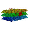
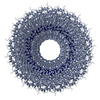
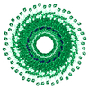
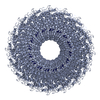
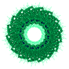
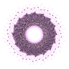
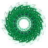
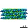
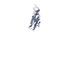
| Title | 4.6 ANGSTROM CRYO-EM RECONSTRUCTION OF TOBACCO MOSAIC VIRUS FROM IMAGES RECORDED AT 300 KEV ON A 4KX4K CCD CAMERA | ||||||
 Components Components |
| ||||||
 Keywords Keywords | VIRUS / SINGLE PARTICLE PROCESSING | ||||||
| Function / homology | Tobacco mosaic virus-like, coat protein / Tobacco mosaic virus-like, coat protein superfamily / Virus coat protein (TMV like) / helical viral capsid / structural molecule activity / RNA / Capsid protein Function and homology information Function and homology information | ||||||
| Biological species |  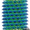 TOBACCO MOSAIC VIRUS TOBACCO MOSAIC VIRUS | ||||||
| Method | ELECTRON MICROSCOPY / helical reconstruction / cryo EM / Resolution: 4.6 Å | ||||||
 Authors Authors | Clare, D.K. / Orlova, E.V. | ||||||
 Citation Citation | Journal: J Struct Biol / Year: 2010 Title: 4.6A Cryo-EM reconstruction of tobacco mosaic virus from images recorded at 300 keV on a 4k x 4k CCD camera. Authors: Daniel K Clare / Elena V Orlova /  Abstract: Tobacco mosaic virus (TMV) is a plant virus with a highly ordered organisation and has been described in three different structural states: As stacked disks without RNA (X-ray crystallography), as a ...Tobacco mosaic virus (TMV) is a plant virus with a highly ordered organisation and has been described in three different structural states: As stacked disks without RNA (X-ray crystallography), as a helical form with RNA (X-ray fibre diffraction) and as a second distinct helical form with RNA (cryo-EM). Here we present a structural analysis of TMV as a test object to assess the quality of cryo-EM images recorded at 300 keV on a CCD camera. The 4.6A TMV structure obtained is consistent with the previous cryo-EM structure and confirms that there is a second helical form of TMV. The structure here also shows that with a similar number of TMV segments an equivalent resolution can be achieved with a 4k CCD camera at 300 keV. | ||||||
| History |
|
- Structure visualization
| Movie |
Movie viewer |
|---|---|
| Structure viewer | Molecule: MolmilJmol/JSmol |
- Downloads & links
-Download
| PDBx/mmCIF format | 2xea.cif.gz | 41.7 KB | Display | PDBx/mmCIF format |
|---|---|---|---|---|
| PDB format | pdb2xea.ent.gz | 27.4 KB | Display | PDB format |
| PDBx/mmJSON format | 2xea.json.gz | Tree view | PDBx/mmJSON format | |
| Others |  Other downloads Other downloads |
-Validation report
| Arichive directory | https://data.pdbj.org/pub/pdb/validation_reports/xe/2xeaftp://data.pdbj.org/pub/pdb/validation_reports/xe/2xea | HTTPS FTP |
|---|
-Related structure data
| Related structure data | 1730MC M: map data used to model this data C: citing same article ( |
|---|---|
| Similar structure data |
-Links
 PDBj
PDBj
- Assembly
| Deposited unit | 
|
|---|---|
| 1 | x 49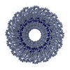
|
| 2 |
|
| 3 | 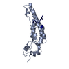
|
| Symmetry | Helical symmetry: (Circular symmetry: 1 / Dyad axis: no / N subunits divisor: 1 / Num. of operations: 49 / Rise per n subunits: 1.408 Å / Rotation per n subunits: 22.032 °) |
-Components
| #1: Protein | Mass: 17505.426 Da / Num. of mol.: 1 / Fragment: CP, RESIDUES 2-159 / Source method: isolated from a natural source / Source: (natural) TOBACCO MOSAIC VIRUS / References: UniProt: Q77LT8 |
|---|---|
| #2: RNA chain | Mass: 958.660 Da / Num. of mol.: 1 / Source method: isolated from a natural source / Source: (natural) TOBACCO MOSAIC VIRUS |
-Experimental details
-Experiment
| Experiment | Method: ELECTRON MICROSCOPY |
|---|---|
| EM experiment | Aggregation state: FILAMENT / 3D reconstruction method: helical reconstruction |
- Sample preparation
| Component | Name: TOBACCO MOSAIC VIRUS / Type: VIRUS |
|---|---|
| Buffer solution | Name: 50 MM TRIS-HCL, 50 MM KCL, 10 MM MGCL2 / pH: 7.4 / Details: 50 MM TRIS-HCL, 50 MM KCL, 10 MM MGCL2 |
| Specimen | Conc.: 3 mg/ml / Embedding applied: NO / Shadowing applied: NO / Staining applied: NO / Vitrification applied: YES |
| Specimen support | Details: HOLEY CARBON |
| Vitrification | Instrument: HOMEMADE PLUNGER / Cryogen name: ETHANE / Details: LIQUID ETHANE |
- Electron microscopy imaging
| Experimental equipment |  Model: Tecnai F30 / Image courtesy: FEI Company |
|---|---|
| Microscopy | Model: FEI TECNAI F30 / Date: Feb 1, 2009 |
| Electron gun | Electron source:  FIELD EMISSION GUN / Accelerating voltage: 300 kV / Illumination mode: FLOOD BEAM FIELD EMISSION GUN / Accelerating voltage: 300 kV / Illumination mode: FLOOD BEAM |
| Electron lens | Mode: BRIGHT FIELD / Nominal magnification: 90000 X / Calibrated magnification: 121000 X / Nominal defocus max: 3000 nm / Nominal defocus min: 900 nm / Cs: 2.3 mm |
| Specimen holder | Temperature: 78 K |
| Image recording | Electron dose: 25 e/Å2 / Film or detector model: GATAN ULTRASCAN 4000 (4k x 4k) |
| Image scans | Num. digital images: 104 |
| Radiation wavelength | Relative weight: 1 |
- Processing
| EM software |
| |||||||||||||||
|---|---|---|---|---|---|---|---|---|---|---|---|---|---|---|---|---|
| CTF correction | Details: EACH PARTICLE WAS FULLY CTF CORRECTED | |||||||||||||||
| 3D reconstruction | Method: PROJECTION MATCHING / Resolution: 4.6 Å / Num. of particles: 5300 / Nominal pixel size: 1.24 Å / Actual pixel size: 1.24 Å Magnification calibration: THE VIRUS DIFFRACTION PATTERN WAS USED TO CALIBRATE THE MICROSCOPE MAGNIFICATION Details: BPRP WAS USED TO RECONSTRUCT THE 3D VOLUME THE PDB 2OM3 WAS DOCKED INTO THE CRYO-EM DENSITY USING CHIMERA. SOME SIDE CHAIN RESIDUES AND THE INNER LOOP OF THE CP WERE REMODELLED USING COOT. ...Details: BPRP WAS USED TO RECONSTRUCT THE 3D VOLUME THE PDB 2OM3 WAS DOCKED INTO THE CRYO-EM DENSITY USING CHIMERA. SOME SIDE CHAIN RESIDUES AND THE INNER LOOP OF THE CP WERE REMODELLED USING COOT. SUBMISSION BASED ON EXPERIMENTAL DATA FROM EMDB EMD-1730 Symmetry type: HELICAL | |||||||||||||||
| Atomic model building | Protocol: RIGID BODY FIT / Space: REAL / Target criteria: Cross-correlation coefficient Details: METHOD--RIGID BODY, REAL SPACE REFINEMENT PROTOCOL--CRYO-EM | |||||||||||||||
| Atomic model building | PDB-ID: 2XEA Accession code: 2XEA / Source name: PDB / Type: experimental model | |||||||||||||||
| Refinement | Highest resolution: 4.6 Å | |||||||||||||||
| Refinement step | Cycle: LAST / Highest resolution: 4.6 Å
|
