[English] 日本語
Yorodumi
- PDB-2wex: Crystal structure of human apoM in complex with glycerol 1- myris... -

+
Open data


ID or keywords:

Loading...

-
Basic information

Entry
Database: PDB / ID: 2wex
TitleCrystal structure of human apoM in complex with glycerol 1- myristic acid
ComponentsAPOLIPOPROTEIN M
KeywordsLIPID TRANSPORT / LIGAND-BINDING SPECIFICITY / GLYCOPROTEIN / MYRISTIC ACID / HDL / LIPOCALIN / TRANSPORT / LIPOPROTEINS
Function / homology
Function and homology information


negative regulation of plasma lipoprotein oxidation / spherical high-density lipoprotein particle / discoidal high-density lipoprotein particle / lipid transporter activity / lipoprotein metabolic process / high-density lipoprotein particle clearance / high-density lipoprotein particle remodeling / very-low-density lipoprotein particle / reverse cholesterol transport / high-density lipoprotein particle assembly ...negative regulation of plasma lipoprotein oxidation / spherical high-density lipoprotein particle / discoidal high-density lipoprotein particle / lipid transporter activity / lipoprotein metabolic process / high-density lipoprotein particle clearance / high-density lipoprotein particle remodeling / very-low-density lipoprotein particle / reverse cholesterol transport / high-density lipoprotein particle assembly / low-density lipoprotein particle / high-density lipoprotein particle / cholesterol efflux / antioxidant activity / Retinoid metabolism and transport / cholesterol homeostasis / phospholipid binding / extracellular region
Similarity search - Function
Apolipoprotein M / ApoM domain / Calycin beta-barrel core domain / Calycin / Lipocalin / Beta Barrel / Mainly Beta
Similarity search - Domain/homology
(2R)-2,3-dihydroxypropyl tetradecanoate / Apolipoprotein M
Similarity search - Component
Biological speciesHOMO SAPIENS (human)
MethodX-RAY DIFFRACTION / SYNCHROTRON / MOLECULAR REPLACEMENT / Resolution: 2 Å
AuthorsSevvana, M. / Ahnstrom, J. / Egerer-Sieber, C. / Dahlback, B. / Muller, Y.A.
CitationJournal: J.Mol.Biol. / Year: 2009
Title: Serendipitous Fatty Acid Binding Reveals the Structural Determinants for Ligand Recognition in Apolipoprotein M.
Authors: Sevvana, M. / Ahnstrom, J. / Egerer-Sieber, C. / Lange, H.A. / Dahlback, B. / Muller, Y.A.
History
DepositionApr 2, 2009Deposition site: PDBE / Processing site: PDBE
Revision 1.0Sep 15, 2009Provider: repository / Type: Initial release
Revision 1.1May 8, 2011Group: Version format compliance
Revision 1.2Jul 13, 2011Group: Version format compliance
Revision 1.3Dec 13, 2023Group: Data collection / Database references ...Data collection / Database references / Derived calculations / Other / Refinement description
Category: chem_comp_atom / chem_comp_bond ...chem_comp_atom / chem_comp_bond / database_2 / pdbx_database_status / pdbx_initial_refinement_model / struct_site
Item: _database_2.pdbx_DOI / _database_2.pdbx_database_accession ..._database_2.pdbx_DOI / _database_2.pdbx_database_accession / _pdbx_database_status.status_code_sf / _struct_site.pdbx_auth_asym_id / _struct_site.pdbx_auth_comp_id / _struct_site.pdbx_auth_seq_id
Remark 650 HELIX DETERMINATION METHOD: AUTHOR PROVIDED.
Remark 700 SHEET THE SHEET STRUCTURE OF THIS MOLECULE IS BIFURCATED. IN ORDER TO REPRESENT THIS FEATURE IN ... SHEET THE SHEET STRUCTURE OF THIS MOLECULE IS BIFURCATED. IN ORDER TO REPRESENT THIS FEATURE IN THE SHEET RECORDS BELOW, TWO SHEETS ARE DEFINED.

-
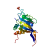
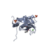
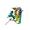
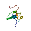
Structure visualization

Structure viewerMolecule:
MolmilJmol/JSmol

Downloads & links

-
Assembly

Deposited unit
A: APOLIPOPROTEIN M
hetero molecules


Theoretical massNumber of molelcules
Total (without water)19,6073
Polymers19,2431
Non-polymers3652
Water1,40578
1


  • Idetical with deposited unit
  • defined by author&software
TypeNameSymmetry operationNumber
identity operation1_555x,y,z1
MethodPQS
Unit cell
Length a, b, c (Å)49.820, 49.820, 145.480
Angle α, β, γ (deg.)90.00, 90.00, 90.00
Int Tables number96
Space group name H-MP43212

-
Components

#1: Protein APOLIPOPROTEIN M / PROTEIN G3A / APO-M / APOM


Mass: 19242.789 Da / Num. of mol.: 1 / Fragment: RESIDUES 22-188
Source method: isolated from a genetically manipulated source
Source: (gene. exp.) HOMO SAPIENS (human) / Production host: ESCHERICHIA COLI (E. coli) / Strain (production host): BL21(DE3) / References: UniProt: O95445
#2: Chemical ChemComp-GYM / (2R)-2,3-dihydroxypropyl tetradecanoate


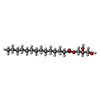
Mass: 302.449 Da / Num. of mol.: 1 / Source method: obtained synthetically / Formula: C17H34O4
#3: Chemical ChemComp-EDO / 1,2-ETHANEDIOL / ETHYLENE GLYCOL


Mass: 62.068 Da / Num. of mol.: 1 / Source method: obtained synthetically / Formula: C2H6O2
#4: Water ChemComp-HOH / water


Mass: 18.015 Da / Num. of mol.: 78 / Source method: isolated from a natural source / Formula: H2O
Sequence detailsRESIDUES 22-188 WITHOUT SIGNAL PEPTIDE HAVE BEEN CLONED

-
Experimental details

-
Experiment

ExperimentMethod: X-RAY DIFFRACTION / Number of used crystals: 1

-
Sample preparation

CrystalDensity Matthews: 2.39 Å3/Da / Density % sol: 47.82 % / Description: NONE
Crystal growpH: 7.2
Details: ABOUT 1-2 MICROL OF PROTEIN (10-16 MG/ML IN 20 MM TRIS-HCL, PH 7.2) WERE MIXED WITH 1-2 MICROL OF 30 % PEG1500 (W/V, IN DEIONISED WATER) AND EQUILIBRATED AGAINST 700 MICROL OF RESERVOIR ...Details: ABOUT 1-2 MICROL OF PROTEIN (10-16 MG/ML IN 20 MM TRIS-HCL, PH 7.2) WERE MIXED WITH 1-2 MICROL OF 30 % PEG1500 (W/V, IN DEIONISED WATER) AND EQUILIBRATED AGAINST 700 MICROL OF RESERVOIR SOLUTION CONTAINING 30 % PEG1500.

-
Data collection

DiffractionMean temperature: 100 K
Diffraction sourceSource: SYNCHROTRON / Site: BESSY / Beamline: 14.1 / Wavelength: 0.91841
DetectorType: RAYONIX / Detector: CCD
RadiationProtocol: SINGLE WAVELENGTH / Monochromatic (M) / Laue (L): M / Scattering type: x-ray
Radiation wavelengthWavelength: 0.91841 Å / Relative weight: 1
ReflectionResolution: 2→20 Å / Num. obs: 13097 / % possible obs: 99.7 % / Observed criterion σ(I): 3 / Redundancy: 8.41 % / Biso Wilson estimate: 42.3 Å2 / Rmerge(I) obs: 0.05 / Net I/σ(I): 23.53
Reflection shellResolution: 2→2.12 Å / Redundancy: 8.49 % / Rmerge(I) obs: 0.54 / Mean I/σ(I) obs: 4.11 / % possible all: 99.7

-
Processing

Software
NameVersionClassification
REFMAC5.2.0019refinement
XDSdata reduction
XSCALEdata scaling
PHASERphasing
RefinementMethod to determine structure: MOLECULAR REPLACEMENT
Starting model: PDB ENTRY 2WEW
Resolution: 2→19 Å / Cor.coef. Fo:Fc: 0.951 / Cor.coef. Fo:Fc free: 0.932 / SU B: 11.127 / SU ML: 0.151 / Cross valid method: THROUGHOUT / ESU R: 0.186 / ESU R Free: 0.173 / Stereochemistry target values: MAXIMUM LIKELIHOOD / Details: HYDROGENS HAVE BEEN ADDED IN THE RIDING POSITIONS.
RfactorNum. reflection% reflectionSelection details
Rfree0.25708 654 5 %RANDOM
Rwork0.20831 ---
obs0.21075 12429 100 %-
Solvent computationIon probe radii: 0.8 Å / Shrinkage radii: 0.8 Å / VDW probe radii: 1.2 Å / Solvent model: MASK
Displacement parametersBiso mean: 19.847 Å2
Baniso -1Baniso -2Baniso -3
1--1.8 Å20 Å20 Å2
2---1.8 Å20 Å2
3---3.61 Å2
Refinement stepCycle: LAST / Resolution: 2→19 Å
ProteinNucleic acidLigandSolventTotal
Num. atoms1253 0 25 78 1356
Refine LS restraints
Refine-IDTypeDev idealDev ideal targetNumber
X-RAY DIFFRACTIONr_bond_refined_d0.0180.0221308
X-RAY DIFFRACTIONr_bond_other_d
X-RAY DIFFRACTIONr_angle_refined_deg1.81.9761761
X-RAY DIFFRACTIONr_angle_other_deg
X-RAY DIFFRACTIONr_dihedral_angle_1_deg7.025156
X-RAY DIFFRACTIONr_dihedral_angle_2_deg37.48724.13858
X-RAY DIFFRACTIONr_dihedral_angle_3_deg18.38115220
X-RAY DIFFRACTIONr_dihedral_angle_4_deg19.787157
X-RAY DIFFRACTIONr_chiral_restr0.1240.2189
X-RAY DIFFRACTIONr_gen_planes_refined0.0080.02975
X-RAY DIFFRACTIONr_gen_planes_other
X-RAY DIFFRACTIONr_nbd_refined0.2330.2490
X-RAY DIFFRACTIONr_nbd_other
X-RAY DIFFRACTIONr_nbtor_refined0.3060.2852
X-RAY DIFFRACTIONr_nbtor_other
X-RAY DIFFRACTIONr_xyhbond_nbd_refined0.1850.269
X-RAY DIFFRACTIONr_xyhbond_nbd_other
X-RAY DIFFRACTIONr_metal_ion_refined
X-RAY DIFFRACTIONr_metal_ion_other
X-RAY DIFFRACTIONr_symmetry_vdw_refined0.2140.241
X-RAY DIFFRACTIONr_symmetry_vdw_other
X-RAY DIFFRACTIONr_symmetry_hbond_refined0.330.27
X-RAY DIFFRACTIONr_symmetry_hbond_other
X-RAY DIFFRACTIONr_symmetry_metal_ion_refined
X-RAY DIFFRACTIONr_symmetry_metal_ion_other
X-RAY DIFFRACTIONr_mcbond_it1.0471.5825
X-RAY DIFFRACTIONr_mcbond_other
X-RAY DIFFRACTIONr_mcangle_it1.52721278
X-RAY DIFFRACTIONr_mcangle_other
X-RAY DIFFRACTIONr_scbond_it2.6063553
X-RAY DIFFRACTIONr_scbond_other
X-RAY DIFFRACTIONr_scangle_it3.6144.5483
X-RAY DIFFRACTIONr_scangle_other
X-RAY DIFFRACTIONr_long_range_B_refined
X-RAY DIFFRACTIONr_long_range_B_other
X-RAY DIFFRACTIONr_rigid_bond_restr
X-RAY DIFFRACTIONr_sphericity_free
X-RAY DIFFRACTIONr_sphericity_bonded
LS refinement shellResolution: 2→2.051 Å / Total num. of bins used: 20
RfactorNum. reflection% reflection
Rfree0.341 46 -
Rwork0.239 873 -
obs--100 %
Refinement TLS params.

Method: refined / Refine-ID: X-RAY DIFFRACTION

IDL112)L122)L132)L222)L232)L332)S11 (Å °)S12 (Å °)S13 (Å °)S21 (Å °)S22 (Å °)S23 (Å °)S31 (Å °)S32 (Å °)S33 (Å °)T112)T122)T132)T222)T232)T332)Origin x (Å)Origin y (Å)Origin z (Å)
129.5951-3.7987.72690.5695-0.62123.68760.85351.7266-2.63790.0104-0.4168-0.57270.69980.4822-0.43670.01430.16320.01560.1868-0.1570.182726.8407-8.497125.864
28.77822.8034-1.07351.25020.11481.42720.02460.58430.1684-0.2173-0.0836-0.31630.0646-0.09350.0590.06470.12470.07520.04540.0713-0.127411.00222.419127.6842
34.1493.02851.11117.28762.8924.82070.04510.67210.3613-0.6295-0.0107-0.10450.0024-0.2462-0.03430.01560.1181-0.04420.00260.0382-0.10932.37366.956727.6652
44.178-1.3631-0.941421.78412.36258.24390.20960.8256-0.0124-1.60880.0741-0.5718-0.8647-0.1064-0.28360.08680.0958-0.06030.00960.0551-0.1652.73664.341121.3593
514.5885-10.0309-2.074218.8791-4.156524.28810.44611.64990.1548-2.2759-0.0442-0.3862-0.5823-0.5138-0.40190.16570.15960.08290.275-0.024-0.18199.109-2.024516.391
610.5745-3.40434.33999.1356-8.494446.70330.08150.506-0.3857-0.3413-0.2035-0.3614-0.20531.91120.122-0.19390.0390.0545-0.0164-0.014-0.177515.0936-2.980625.1634
78.814-0.6549-1.13042.87660.17672.5658-0.03930.46080.3678-0.2749-0.0547-0.37830.0040.25070.094-0.09850.05410.0253-0.03980.05710.012317.49592.623330.3081
87.4368-4.3868-2.29925.05372.26664.30950.0607-0.0871.0329-0.02060.2244-0.3632-0.15220.2938-0.2851-0.11240.0102-0.0155-0.12620.0851-0.0313.260110.138134.4255
Refinement TLS group
IDRefine-IDRefine TLS-IDAuth asym-IDAuth seq-ID
1X-RAY DIFFRACTION1A18 - 31
2X-RAY DIFFRACTION2A36 - 63
3X-RAY DIFFRACTION3A64 - 92
4X-RAY DIFFRACTION4A93 - 108
5X-RAY DIFFRACTION5A109 - 113
6X-RAY DIFFRACTION6A119 - 129
7X-RAY DIFFRACTION7A130 - 167
8X-RAY DIFFRACTION8A168 - 185

+
About Yorodumi

-
News

-
Feb 9, 2022. New format data for meta-information of EMDB entries

New format data for meta-information of EMDB entries

  • Version 3 of the EMDB header file is now the official format.
  • The previous official version 1.9 will be removed from the archive.

Related info.:EMDB header

External links:wwPDB to switch to version 3 of the EMDB data model

-
Aug 12, 2020. Covid-19 info

Covid-19 info

URL: https://pdbjlvh1.pdbj.org/emnavi/covid19.php

New page: Covid-19 featured information page in EM Navigator.

Related info.:Covid-19 info / Mar 5, 2020. Novel coronavirus structure data

+
Mar 5, 2020. Novel coronavirus structure data

Novel coronavirus structure data

Related info.:Yorodumi Speices / Aug 12, 2020. Covid-19 info

External links:COVID-19 featured content - PDBj / Molecule of the Month (242):Coronavirus Proteases

+
Jan 31, 2019. EMDB accession codes are about to change! (news from PDBe EMDB page)

EMDB accession codes are about to change! (news from PDBe EMDB page)

  • The allocation of 4 digits for EMDB accession codes will soon come to an end. Whilst these codes will remain in use, new EMDB accession codes will include an additional digit and will expand incrementally as the available range of codes is exhausted. The current 4-digit format prefixed with “EMD-” (i.e. EMD-XXXX) will advance to a 5-digit format (i.e. EMD-XXXXX), and so on. It is currently estimated that the 4-digit codes will be depleted around Spring 2019, at which point the 5-digit format will come into force.
  • The EM Navigator/Yorodumi systems omit the EMD- prefix.

Related info.:Q: What is EMD? / ID/Accession-code notation in Yorodumi/EM Navigator

External links:EMDB Accession Codes are Changing Soon! / Contact to PDBj

+
Jul 12, 2017. Major update of PDB

Major update of PDB

  • wwPDB released updated PDB data conforming to the new PDBx/mmCIF dictionary.
  • This is a major update changing the version number from 4 to 5, and with Remediation, in which all the entries are updated.
  • In this update, many items about electron microscopy experimental information are reorganized (e.g. em_software).
  • Now, EM Navigator and Yorodumi are based on the updated data.

External links:wwPDB Remediation / Enriched Model Files Conforming to OneDep Data Standards Now Available in the PDB FTP Archive

-
Yorodumi

Thousand views of thousand structures

  • Yorodumi is a browser for structure data from EMDB, PDB, SASBDB, etc.
  • This page is also the successor to EM Navigator detail page, and also detail information page/front-end page for Omokage search.
  • The word "yorodu" (or yorozu) is an old Japanese word meaning "ten thousand". "mi" (miru) is to see.

Related info.:EMDB / PDB / SASBDB / Comparison of 3 databanks / Yorodumi Search / Aug 31, 2016. New EM Navigator & Yorodumi / Yorodumi Papers / Jmol/JSmol / Function and homology information / Changes in new EM Navigator and Yorodumi

Read more