 Movie
Movie Controller

[English] 日本語
 Yorodumi
- PDB-2tpl: TYROSINE PHENOL-LYASE FROM CITROBACTER INTERMEDIUS COMPLEX WITH 3... -
+ Open data

- Basic information
| Entry | Database: PDB / ID: 2tpl | ||||||
|---|---|---|---|---|---|---|---|
| Title | TYROSINE PHENOL-LYASE FROM CITROBACTER INTERMEDIUS COMPLEX WITH 3-(4'-HYDROXYPHENYL)PROPIONIC ACID, PYRIDOXAL-5'-PHOSPHATE AND CS+ ION | ||||||
 Components Components | TYROSINE PHENOL-LYASE | ||||||
 Keywords Keywords | LYASE / PLP-DEPENDENT ENZYME / PYRIDOXAL PHOSPHATE | ||||||
| Function / homology |  Function and homology information Function and homology information | ||||||
| Biological species |  Citrobacter freundii (bacteria) Citrobacter freundii (bacteria) | ||||||
| Method |  X-RAY DIFFRACTION / SYNCHROTRON / MOLECULAR REPLACEMENT / Resolution: 2.5 Å X-RAY DIFFRACTION / SYNCHROTRON / MOLECULAR REPLACEMENT / Resolution: 2.5 Å | ||||||
 Authors Authors | Antson, A.A. / Demidkina, T.V. / Wilson, K.S. | ||||||
 Citation Citation | Journal: Biochemistry / Year: 1997 Title: The crystal structure of Citrobacter freundii tyrosine phenol-lyase complexed with 3-(4'-hydroxyphenyl)propionic acid, together with site-directed mutagenesis and kinetic analysis, ...Title: The crystal structure of Citrobacter freundii tyrosine phenol-lyase complexed with 3-(4'-hydroxyphenyl)propionic acid, together with site-directed mutagenesis and kinetic analysis, demonstrates that arginine 381 is required for substrate specificity. Authors: Sundararaju, B. / Antson, A.A. / Phillips, R.S. / Demidkina, T.V. / Barbolina, M.V. / Gollnick, P. / Dodson, G.G. / Wilson, K.S. | ||||||
| History |
|
- Structure visualization
| Structure viewer | Molecule: MolmilJmol/JSmol |
|---|
- Downloads & links
-Download
| PDBx/mmCIF format | 2tpl.cif.gz | 194.7 KB | Display | PDBx/mmCIF format |
|---|---|---|---|---|
| PDB format | pdb2tpl.ent.gz | 154.1 KB | Display | PDB format |
| PDBx/mmJSON format | 2tpl.json.gz | Tree view | PDBx/mmJSON format | |
| Others |  Other downloads Other downloads |
-Validation report
| Arichive directory | https://data.pdbj.org/pub/pdb/validation_reports/tp/2tplftp://data.pdbj.org/pub/pdb/validation_reports/tp/2tpl | HTTPS FTP |
|---|
-Related structure data
| Similar structure data |
|---|

-Links
 PDBj
PDBj- Assembly
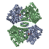
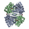
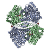
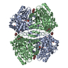
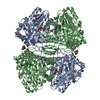
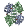
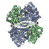
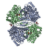
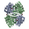
| Deposited unit | 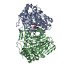
| ||||||||
|---|---|---|---|---|---|---|---|---|---|
| 1 | 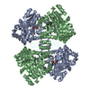
| ||||||||
| Unit cell |
| ||||||||
| Noncrystallographic symmetry (NCS) | NCS oper: (Code: given Matrix: (0.978, -0.208, -0.004), Vector: |
-Components
| #1: Protein | Mass: 51736.871 Da / Num. of mol.: 2 / Source method: isolated from a natural source / Source: (natural) Citrobacter freundii (bacteria) / References: UniProt: P31013, tyrosine phenol-lyase#2: Chemical |   Mass: 132.905 Da / Num. of mol.: 2 / Source method: obtained synthetically / Formula: Cs Mass: 132.905 Da / Num. of mol.: 2 / Source method: obtained synthetically / Formula: Cs#3: Chemical | ChemComp-HPP / | 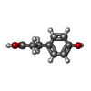  Mass: 166.174 Da / Num. of mol.: 1 / Source method: obtained synthetically / Formula: C9H10O3 Mass: 166.174 Da / Num. of mol.: 1 / Source method: obtained synthetically / Formula: C9H10O3#4: Water | ChemComp-HOH / |  Mass: 18.015 Da / Num. of mol.: 252 / Source method: isolated from a natural source / Formula: H2O Mass: 18.015 Da / Num. of mol.: 252 / Source method: isolated from a natural source / Formula: H2O |
|---|
-Experimental details
-Experiment
| Experiment | Method: X-RAY DIFFRACTION / Number of used crystals: 3 |
|---|
- Sample preparation
| Crystal | Density Matthews: 2.8 Å3/Da / Density % sol: 56 % | ||||||||||||||||||||||||||||||||||||||||||||||||||||||||
|---|---|---|---|---|---|---|---|---|---|---|---|---|---|---|---|---|---|---|---|---|---|---|---|---|---|---|---|---|---|---|---|---|---|---|---|---|---|---|---|---|---|---|---|---|---|---|---|---|---|---|---|---|---|---|---|---|---|
| Crystal grow | pH: 8 Details: 30-45% PEG 5000 MONOMETHYL ETHER, 0.4 M CSCL, 50 MM TRIETHANOLAMINE BUFFER PH 8.0, 1 MM DTT, 0.2 MM PLP, 50 MM HPPA | ||||||||||||||||||||||||||||||||||||||||||||||||||||||||
| Crystal grow | *PLUS Method: vapor diffusion, hanging dropDetails: drop solution is half diluted with a reservoir solution | ||||||||||||||||||||||||||||||||||||||||||||||||||||||||
| Components of the solutions | *PLUS
|
-Data collection
| Diffraction | Mean temperature: 293 K |
|---|---|
| Diffraction source | Source: SYNCHROTRON / Site: SRS  / Beamline: PX9.5 / Wavelength: 0.9177 / Beamline: PX9.5 / Wavelength: 0.9177 |
| Detector | Type: MARRESEARCH / Detector: IMAGE PLATE / Date: Mar 1, 1994 / Details: PT-COATED TOROIDAL MIRROR |
| Radiation | Monochromator: SI(111) / Monochromatic (M) / Laue (L): M / Scattering type: x-ray |
| Radiation wavelength | Wavelength: 0.9177 Å / Relative weight: 1 |
| Reflection | Resolution: 2.5→15 Å / Num. obs: 33108 / % possible obs: 81 % / Observed criterion σ(I): 0 / Redundancy: 2.7 % / Rmerge(I) obs: 0.139 / Net I/σ(I): 4.7 |
| Reflection shell | Resolution: 2.5→2.6 Å / Redundancy: 1.7 % / Rmerge(I) obs: 0.361 / Mean I/σ(I) obs: 1.9 / % possible all: 50 |
| Reflection shell | *PLUS % possible obs: 50.3 % |
- Processing
| Software |
| ||||||||||||||||||||||||||||||||
|---|---|---|---|---|---|---|---|---|---|---|---|---|---|---|---|---|---|---|---|---|---|---|---|---|---|---|---|---|---|---|---|---|---|
| Refinement | Method to determine structure: MOLECULAR REPLACEMENT Starting model: TYROSINE PHENOL-LYASE HOLOENZYME Resolution: 2.5→15 Å / Cross valid method: THROUGHOUT / σ(F): 0
| ||||||||||||||||||||||||||||||||
| Refinement step | Cycle: LAST / Resolution: 2.5→15 Å
| ||||||||||||||||||||||||||||||||
| Software | *PLUS Name: REFMAC / Classification: refinement | ||||||||||||||||||||||||||||||||
| Refinement | *PLUS Num. reflection Rfree: 957 | ||||||||||||||||||||||||||||||||
| Solvent computation | *PLUS | ||||||||||||||||||||||||||||||||
| Displacement parameters | *PLUS Biso mean: 29.4 Å2 | ||||||||||||||||||||||||||||||||
| Refine LS restraints | *PLUS
|
