[English] 日本語
 Yorodumi
- PDB-2gst: STRUCTURE OF THE XENOBIOTIC SUBSTRATE BINDING SITE OF A GLUTATHIO... -
+ Open data
- Basic information
| Entry | Database: PDB / ID: 2gst | ||||||
|---|---|---|---|---|---|---|---|
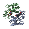
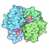
| Title | STRUCTURE OF THE XENOBIOTIC SUBSTRATE BINDING SITE OF A GLUTATHIONE S-TRANSFERASE AS REVEALED BY X-RAY CRYSTALLOGRAPHIC ANALYSIS OF PRODUCT COMPLEXES WITH THE DIASTEREOMERS OF 9-(S-GLUTATHIONYL)-10-HYDROXY-9, 10-DIHYDROPHENANTHRENE | ||||||
 Components Components | GLUTATHIONE S-TRANSFERASE | ||||||
 Keywords Keywords | GLUTATHIONE TRANSFERASE | ||||||
| Function / homology |  Function and homology information Function and homology informationGlutathione conjugation / nitrobenzene metabolic process / cellular detoxification of nitrogen compound / hepoxilin biosynthetic process / glutathione derivative biosynthetic process / glutathione binding / response to metal ion / prostaglandin metabolic process / glutathione transferase / nickel cation binding ...Glutathione conjugation / nitrobenzene metabolic process / cellular detoxification of nitrogen compound / hepoxilin biosynthetic process / glutathione derivative biosynthetic process / glutathione binding / response to metal ion / prostaglandin metabolic process / glutathione transferase / nickel cation binding / glutathione transferase activity / response to amino acid / xenobiotic catabolic process / response to axon injury / steroid binding / glutathione metabolic process / response to lead ion / cellular response to xenobiotic stimulus / sensory perception of smell / response to ethanol / protein kinase binding / enzyme binding / protein homodimerization activity / protein-containing complex / mitochondrion / extracellular region / identical protein binding / cytosol / cytoplasm Similarity search - Function | ||||||
| Biological species |  | ||||||
| Method |  X-RAY DIFFRACTION / Resolution: 1.8 Å X-RAY DIFFRACTION / Resolution: 1.8 Å | ||||||
 Authors Authors | Ji, X. / Armstrong, R.N. / Gilliland, G.L. | ||||||
 Citation Citation |  Journal: Biochemistry / Year: 1994 Journal: Biochemistry / Year: 1994Title: Structure and function of the xenobiotic substrate binding site of a glutathione S-transferase as revealed by X-ray crystallographic analysis of product complexes with the diastereomers of 9- ...Title: Structure and function of the xenobiotic substrate binding site of a glutathione S-transferase as revealed by X-ray crystallographic analysis of product complexes with the diastereomers of 9-(S-glutathionyl)-10-hydroxy-9,10-dihydrophenanthrene. Authors: Ji, X. / Johnson, W.W. / Sesay, M.A. / Dickert, L. / Prasad, S.M. / Ammon, H.L. / Armstrong, R.N. / Gilliland, G.L. #1:  Journal: J.Biol.Chem. / Year: 1993 Journal: J.Biol.Chem. / Year: 1993Title: Tyrosine 115 Participates Both in Chemical and Physical Steps of the Catalytic Mechanism of a Glutathione S-Transferase Authors: Johnson, W.W. / Liu, S. / Ji, X. / Gilliland, G.L. / Armstrong, R.N. #2:  Journal: Biochemistry / Year: 1992 Journal: Biochemistry / Year: 1992Title: The Three-Dimensional Structure of a Glutathione S-Transferase from the Mu Gene Class. Structural Analysis of the Binary Complex of Isoenzyme 3-3 and Glutothione at 2.2 Angstroms Resolution Authors: Ji, X. / Zhang, P. / Armstrong, R.N. / Gilliland, G.L. #3:  Journal: J.Biol.Chem. / Year: 1992 Journal: J.Biol.Chem. / Year: 1992Title: Contribution of Tyrosine 6 to the Catalytic Mechanism of Isoenzyme 3-3 of Glutathione S-Transferase Authors: Liu, S. / Zhang, P. / Ji, X. / Johnson, W.W. / Gilliland, G.L. / Armstrong, R.N. | ||||||
| History |
| ||||||
| Remark 650 | HELIX H5A AND H5B OF THE HELIX MAY BE CONSIDERED AS A SINGLE LONG HELIX WHICH BENDS BY ABOUT 35 DEGREES. |
- Structure visualization
| Structure viewer | Molecule:  Molmil Molmil Jmol/JSmol Jmol/JSmol |
|---|
- Downloads & links
- Download
| PDBx/mmCIF format |  2gst.cif.gz 2gst.cif.gz | 115.4 KB | Display |  PDBx/mmCIF format PDBx/mmCIF format |
|---|---|---|---|---|
| PDB format |  pdb2gst.ent.gz pdb2gst.ent.gz | 88.7 KB | Display |  PDB format PDB format |
| PDBx/mmJSON format |  2gst.json.gz 2gst.json.gz | Tree view |  PDBx/mmJSON format PDBx/mmJSON format | |
| Others |  Other downloads Other downloads |
-Validation report
| Summary document |  2gst_validation.pdf.gz 2gst_validation.pdf.gz | 531.8 KB | Display |  wwPDB validaton report wwPDB validaton report |
|---|---|---|---|---|
| Full document |  2gst_full_validation.pdf.gz 2gst_full_validation.pdf.gz | 552.2 KB | Display | |
| Data in XML |  2gst_validation.xml.gz 2gst_validation.xml.gz | 13.9 KB | Display | |
| Data in CIF |  2gst_validation.cif.gz 2gst_validation.cif.gz | 22 KB | Display | |
| Arichive directory |  https://data.pdbj.org/pub/pdb/validation_reports/gs/2gst https://data.pdbj.org/pub/pdb/validation_reports/gs/2gst ftp://data.pdbj.org/pub/pdb/validation_reports/gs/2gst ftp://data.pdbj.org/pub/pdb/validation_reports/gs/2gst | HTTPS FTP |
-Related structure data
- Links
Links
- Assembly
| Deposited unit | 
| |||||||||
|---|---|---|---|---|---|---|---|---|---|---|
| 1 |
| |||||||||
| Unit cell |
| |||||||||
| Atom site foot note | 1: RESIDUES 38, 60, AND 206 OF BOTH CHAINS ARE CIS PROLINES. | |||||||||
| Components on special symmetry positions |
| |||||||||
| Noncrystallographic symmetry (NCS) | NCS oper: (Code: given Matrix: (0.8188, -0.1067, 0.5641), Vector: Details | THE TRANSFORMATION PRESENTED ON *MTRIX* RECORDS BELOW WILL YIELD APPROXIMATE COORDINATES FOR CHAIN *B* WHEN APPLIED TO CHAIN *A*, WHICH CORRESPONDS TO A ROTATION OF 179.749 DEGREES AROUND THE DIRECTION 0.9536 -0.0553 0.2959. | |
- Components
| #1: Protein | Mass: 25818.791 Da / Num. of mol.: 2 Source method: isolated from a genetically manipulated source Source: (gene. exp.)  #2: Chemical | #3: Chemical | #4: Water | ChemComp-HOH / | |
|---|
-Experimental details
-Experiment
| Experiment | Method:  X-RAY DIFFRACTION X-RAY DIFFRACTION |
|---|
- Sample preparation
| Crystal | Density Matthews: 2.32 Å3/Da / Density % sol: 46.9 % |
|---|
-Data collection
| Reflection | *PLUS Highest resolution: 1.8 Å / Num. all: 168166 / Num. obs: 42010 / Observed criterion σ(I): 2 / Num. measured all: 40244 / Rmerge F obs: 0.0732 |
|---|
- Processing
| Software | Name: GPRLSA / Classification: refinement | |||||||||||||||||||||||||||||||||||||||||||||||||||||||||||||||
|---|---|---|---|---|---|---|---|---|---|---|---|---|---|---|---|---|---|---|---|---|---|---|---|---|---|---|---|---|---|---|---|---|---|---|---|---|---|---|---|---|---|---|---|---|---|---|---|---|---|---|---|---|---|---|---|---|---|---|---|---|---|---|---|---|
| Refinement | Rfactor obs: 0.16 / Highest resolution: 1.8 Å | |||||||||||||||||||||||||||||||||||||||||||||||||||||||||||||||
| Refinement step | Cycle: LAST / Highest resolution: 1.8 Å
| |||||||||||||||||||||||||||||||||||||||||||||||||||||||||||||||
| Refine LS restraints |
| |||||||||||||||||||||||||||||||||||||||||||||||||||||||||||||||
| Refinement | *PLUS Highest resolution: 1.8 Å / Lowest resolution: 6 Å / Num. reflection obs: 35708 / σ(I): 2 / Rfactor obs: 0.16 | |||||||||||||||||||||||||||||||||||||||||||||||||||||||||||||||
| Solvent computation | *PLUS | |||||||||||||||||||||||||||||||||||||||||||||||||||||||||||||||
| Displacement parameters | *PLUS | |||||||||||||||||||||||||||||||||||||||||||||||||||||||||||||||
| Refine LS restraints | *PLUS
|
 Movie
Movie Controller


 PDBj
PDBj


