 Movie
Movie Controller

[English] 日本語
 Yorodumi
- PDB-2fzc: The Structure of Wild-Type E. Coli Aspartate Transcarbamoylase in... -
+ Open data

- Basic information
| Entry | Database: PDB / ID: 2fzc | ||||||
|---|---|---|---|---|---|---|---|
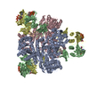
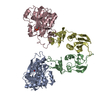
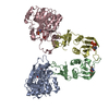
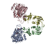
| Title | The Structure of Wild-Type E. Coli Aspartate Transcarbamoylase in Complex with Novel T State Inhibitors at 2.10 Resolution | ||||||
 Components Components | (Aspartate carbamoyltransferase ...) x 2 | ||||||
 Keywords Keywords | TRANSFERASE / Inhibitors / allosteric transition / Xray Structures | ||||||
| Function / homology |  Function and homology information Function and homology informationaspartate carbamoyltransferase complex / pyrimidine nucleotide biosynthetic process / aspartate carbamoyltransferase / aspartate carbamoyltransferase activity / L-glutamine metabolic process / amino acid binding / protein homotrimerization / 'de novo' UMP biosynthetic process / 'de novo' pyrimidine nucleobase biosynthetic process / zinc ion binding ...aspartate carbamoyltransferase complex / pyrimidine nucleotide biosynthetic process / aspartate carbamoyltransferase / aspartate carbamoyltransferase activity / L-glutamine metabolic process / amino acid binding / protein homotrimerization / 'de novo' UMP biosynthetic process / 'de novo' pyrimidine nucleobase biosynthetic process / zinc ion binding / identical protein binding / cytosol / cytoplasm Similarity search - Function | ||||||
| Biological species |  | ||||||
| Method |  X-RAY DIFFRACTION / MOLECULAR REPLACEMENT / Resolution: 2.1 Å X-RAY DIFFRACTION / MOLECULAR REPLACEMENT / Resolution: 2.1 Å | ||||||
 Authors Authors | Heng, S. / Stieglitz, K.A. / Eldo, J. / Xia, J. / Cardia, J.P. / Kantrowitz, E.R. | ||||||
 Citation Citation | Journal: Biochemistry / Year: 2006 Title: T-state Inhibitors of E. coli Aspartate Transcarbamoylase that Prevent the Allosteric Transition. Authors: Heng, S. / Stieglitz, K.A. / Eldo, J. / Xia, J. / Cardia, J.P. / Kantrowitz, E.R. | ||||||
| History |
|
- Structure visualization
| Structure viewer | Molecule: MolmilJmol/JSmol |
|---|
- Downloads & links
-Download
| PDBx/mmCIF format | 2fzc.cif.gz | 211.8 KB | Display | PDBx/mmCIF format |
|---|---|---|---|---|
| PDB format | pdb2fzc.ent.gz | 167.1 KB | Display | PDB format |
| PDBx/mmJSON format | 2fzc.json.gz | Tree view | PDBx/mmJSON format | |
| Others |  Other downloads Other downloads |
-Validation report
| Arichive directory | https://data.pdbj.org/pub/pdb/validation_reports/fz/2fzcftp://data.pdbj.org/pub/pdb/validation_reports/fz/2fzc | HTTPS FTP |
|---|
-Related structure data
| Related structure data |  2fzgC  2fzkC  1za1S S: Starting model for refinement C: citing same article ( |
|---|---|
| Similar structure data |


-Links
 PDBj
PDBj
- Assembly
| Deposited unit | 
| ||||||||
|---|---|---|---|---|---|---|---|---|---|
| 1 | 
| ||||||||
| Unit cell |
|
-Components
-Aspartate carbamoyltransferase ... , 2 types, 4 molecules ACBD
| #1: Protein | Mass: 34337.105 Da / Num. of mol.: 2 Source method: isolated from a genetically manipulated source Source: (gene. exp.) #2: Protein | Mass: 17143.625 Da / Num. of mol.: 2 Source method: isolated from a genetically manipulated source Source: (gene. exp.) |
|---|
-Non-polymers , 4 types, 773 molecules 


| #3: Chemical |  Mass: 304.131 Da / Num. of mol.: 2 / Source method: obtained synthetically / Formula: C6H14N2O8P2 Mass: 304.131 Da / Num. of mol.: 2 / Source method: obtained synthetically / Formula: C6H14N2O8P2#4: Chemical |  Mass: 65.409 Da / Num. of mol.: 2 / Source method: obtained synthetically / Formula: Zn Mass: 65.409 Da / Num. of mol.: 2 / Source method: obtained synthetically / Formula: Zn#5: Chemical |  Mass: 483.156 Da / Num. of mol.: 2 / Source method: obtained synthetically / Formula: C9H16N3O14P3 Mass: 483.156 Da / Num. of mol.: 2 / Source method: obtained synthetically / Formula: C9H16N3O14P3#6: Water | ChemComp-HOH / | Mass: 18.015 Da / Num. of mol.: 767 / Source method: isolated from a natural source / Formula: H2O |
|---|
-Experimental details
-Experiment
| Experiment | Method: X-RAY DIFFRACTION / Number of used crystals: 1 |
|---|
- Sample preparation
| Crystal | Density Matthews: 2.89 Å3/Da / Density % sol: 57.41 % |
|---|---|
| Crystal grow | Temperature: 298 K / Method: vapor diffusion, hanging drop / pH: 5.7 Details: ATCase holoenzyme was crystallized by microdialysis, using 50 L wells. The enzyme solution, at ~18 mg/mL, was dialyzed against a solution of 40 mM citric acid, 3 mM sodium azide, 1 mM 2- ...Details: ATCase holoenzyme was crystallized by microdialysis, using 50 L wells. The enzyme solution, at ~18 mg/mL, was dialyzed against a solution of 40 mM citric acid, 3 mM sodium azide, 1 mM 2-mercaptoethanol, 1 mM cytidine 5 -triphosphate, 0.2 mM EDTA (pH 5.7) , VAPOR DIFFUSION, HANGING DROP, temperature 298K |
-Data collection
| Diffraction | Mean temperature: 110 K |
|---|---|
| Diffraction source | Source: ROTATING ANODE / Type: RIGAKU RU200 / Wavelength: 1.5418 Å |
| Detector | Type: RIGAKU RAXIS IV / Detector: IMAGE PLATE / Date: Jan 1, 2005 |
| Radiation | Protocol: SINGLE WAVELENGTH / Monochromatic (M) / Laue (L): M / Scattering type: x-ray |
| Radiation wavelength | Wavelength: 1.5418 Å / Relative weight: 1 |
| Reflection | Resolution: 2→27.35 Å / Num. all: 80677 / Num. obs: 80677 / % possible obs: 99.9 % / Observed criterion σ(F): 0 / Observed criterion σ(I): 0 / Redundancy: 5.31 % / Rmerge(I) obs: 0.058 / Net I/σ(I): 13.5 |
| Reflection shell | Resolution: 2→2.07 Å / Redundancy: 5.13 % / Rmerge(I) obs: 0.362 / Mean I/σ(I) obs: 4.3 / Num. unique all: 8005 / % possible all: 99.5 |
- Processing
| Software |
| |||||||||||||||||||||
|---|---|---|---|---|---|---|---|---|---|---|---|---|---|---|---|---|---|---|---|---|---|---|
| Refinement | Method to determine structure: MOLECULAR REPLACEMENT Starting model: PDB entry 1ZA1 Resolution: 2.1→27.35 Å / Cross valid method: THROUGHOUT / σ(F): 0 / Stereochemistry target values: Engh & Huber
| |||||||||||||||||||||
| Refinement step | Cycle: LAST / Resolution: 2.1→27.35 Å
|
