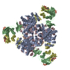
 Movie
Movie Controller

[English] 日本語
 Yorodumi
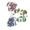
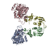
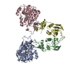
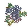
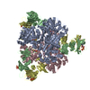
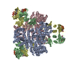
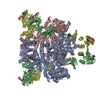
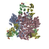
- PDB-1tu0: Aspartate Transcarbamoylase Catalytic Chain Mutant E50A Complex w... -
+ Open data

- Basic information
| Entry | Database: PDB / ID: 1tu0 | ||||||
|---|---|---|---|---|---|---|---|
| Title | Aspartate Transcarbamoylase Catalytic Chain Mutant E50A Complex with Phosphonoacetamide | ||||||
 Components Components |
| ||||||
 Keywords Keywords | HYDROLASE/HYDROLASE REGULATOR / protein structure-function / site-specific mutagenesis / domain closure / allosteric transition / HYDROLASE-HYDROLASE REGULATOR COMPLEX | ||||||
| Function / homology |  Function and homology information Function and homology informationaspartate carbamoyltransferase complex / pyrimidine nucleotide biosynthetic process / aspartate carbamoyltransferase / aspartate carbamoyltransferase activity / L-glutamine metabolic process / amino acid metabolic process / amino acid binding / protein homotrimerization / 'de novo' UMP biosynthetic process / 'de novo' pyrimidine nucleobase biosynthetic process ...aspartate carbamoyltransferase complex / pyrimidine nucleotide biosynthetic process / aspartate carbamoyltransferase / aspartate carbamoyltransferase activity / L-glutamine metabolic process / amino acid metabolic process / amino acid binding / protein homotrimerization / 'de novo' UMP biosynthetic process / 'de novo' pyrimidine nucleobase biosynthetic process / zinc ion binding / identical protein binding / cytosol / cytoplasm Similarity search - Function | ||||||
| Biological species |  | ||||||
| Method |  X-RAY DIFFRACTION / MOLECULAR REPLACEMENT / Resolution: 2.55 Å X-RAY DIFFRACTION / MOLECULAR REPLACEMENT / Resolution: 2.55 Å | ||||||
 Authors Authors | Stieglitz, K. / Stec, B. / Baker, D.P. / Kantrowitz, E.R. | ||||||
 Citation Citation | Journal: J.Mol.Biol. / Year: 2004 Title: Monitoring the Transition from the T to the R State in E.coli Aspartate Transcarbamoylase by X-ray Crystallography: Crystal Structures of the E50A Mutant Enzyme in Four Distinct Allosteric States. Authors: Stieglitz, K. / Stec, B. / Baker, D.P. / Kantrowitz, E.R. | ||||||
| History |
|
- Structure visualization
| Structure viewer | Molecule: MolmilJmol/JSmol |
|---|
- Downloads & links
-Download
| PDBx/mmCIF format | 1tu0.cif.gz | 198.2 KB | Display | PDBx/mmCIF format |
|---|---|---|---|---|
| PDB format | pdb1tu0.ent.gz | 158.6 KB | Display | PDB format |
| PDBx/mmJSON format | 1tu0.json.gz | Tree view | PDBx/mmJSON format | |
| Others |  Other downloads Other downloads |
-Validation report
| Arichive directory | https://data.pdbj.org/pub/pdb/validation_reports/tu/1tu0ftp://data.pdbj.org/pub/pdb/validation_reports/tu/1tu0 | HTTPS FTP |
|---|
-Related structure data


-Links
 PDBj
PDBj
- Assembly
| Deposited unit | 
| |||||||||
|---|---|---|---|---|---|---|---|---|---|---|
| 1 | 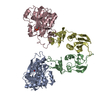
| |||||||||
| Unit cell |
| |||||||||
| Components on special symmetry positions |
|
-Components
| #1: Protein | Mass: 34279.074 Da / Num. of mol.: 2 / Mutation: E50A Source method: isolated from a genetically manipulated source Source: (gene. exp.) #2: Protein | Mass: 17143.625 Da / Num. of mol.: 2 Source method: isolated from a genetically manipulated source Source: (gene. exp.) #3: Chemical |   Mass: 139.047 Da / Num. of mol.: 2 / Source method: obtained synthetically / Formula: C2H6NO4P Mass: 139.047 Da / Num. of mol.: 2 / Source method: obtained synthetically / Formula: C2H6NO4P#4: Chemical |   Mass: 65.409 Da / Num. of mol.: 2 / Source method: obtained synthetically / Formula: Zn Mass: 65.409 Da / Num. of mol.: 2 / Source method: obtained synthetically / Formula: Zn#5: Water | ChemComp-HOH / |  Mass: 18.015 Da / Num. of mol.: 354 / Source method: isolated from a natural source / Formula: H2O Mass: 18.015 Da / Num. of mol.: 354 / Source method: isolated from a natural source / Formula: H2O |
|---|
-Experimental details
-Experiment
| Experiment | Method: X-RAY DIFFRACTION / Number of used crystals: 1 |
|---|
- Sample preparation
| Crystal | Density Matthews: 2.23 Å3/Da / Density % sol: 44.9 % |
|---|---|
| Crystal grow | Temperature: 295 K / pH: 6 Details: The Glu50Ala mutant was crystallized in 100 mM sodium citrate, 1 mM 2-mercaptoethanol, and 15% PEG 8000 pH 7.0. Before mounting, 50 mM PAM was added and the crystal soaked, pH 6.0, ...Details: The Glu50Ala mutant was crystallized in 100 mM sodium citrate, 1 mM 2-mercaptoethanol, and 15% PEG 8000 pH 7.0. Before mounting, 50 mM PAM was added and the crystal soaked, pH 6.0, MICRODIALYSIS, temperature 295K, pH 6.00 |
-Data collection
| Diffraction | Mean temperature: 298 K |
|---|---|
| Diffraction source | Source: ROTATING ANODE / Type: RIGAKU RU200 / Wavelength: 1.5418 |
| Detector | Type: UCSD MARK III / Detector: AREA DETECTOR / Date: Mar 31, 1998 |
| Radiation | Monochromator: GRAPHITE / Protocol: SINGLE WAVELENGTH / Monochromatic (M) / Laue (L): M / Scattering type: x-ray |
| Radiation wavelength | Wavelength: 1.5418 Å / Relative weight: 1 |
| Reflection | Resolution: 2.55→30 Å / Num. obs: 37788 / % possible obs: 94 % / Observed criterion σ(I): 2 / Redundancy: 3.8 % / Rmerge(I) obs: 0.085 / Net I/σ(I): 7.37 |
| Reflection shell | Resolution: 2.55→2.6 Å / Redundancy: 3.5 % / Rmerge(I) obs: 0.36 / Mean I/σ(I) obs: 1.32 / % possible all: 89.53 |
- Processing
| Software |
| |||||||||||||||||||||||||||||||||
|---|---|---|---|---|---|---|---|---|---|---|---|---|---|---|---|---|---|---|---|---|---|---|---|---|---|---|---|---|---|---|---|---|---|---|
| Refinement | Method to determine structure: MOLECULAR REPLACEMENT Starting model: UNPUBLISHED Resolution: 2.55→30 Å / Num. parameters: 30387 / Num. restraintsaints: 29984 / Cross valid method: THROUGHOUT / σ(F): 0 / Stereochemistry target values: ENGH AND HUBER Details: GEOMETRY OF STRUCTURES WAS CHECKED AND CORRECTED IN CNS. FINAL RUNS FOR THIS ENTRY WERE CHECKED AGAINST SFCHECK YIELDED R-FACTOR AND R-FREE.
| |||||||||||||||||||||||||||||||||
| Refine analyze | Num. disordered residues: 0 / Occupancy sum hydrogen: 0 / Occupancy sum non hydrogen: 7585.11 | |||||||||||||||||||||||||||||||||
| Refinement step | Cycle: LAST / Resolution: 2.55→30 Å
| |||||||||||||||||||||||||||||||||
| Refine LS restraints |
|
