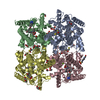
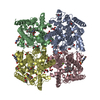
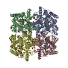
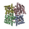
Entry Database : PDB / ID : 2fm5Title Crystal structure of PDE4D2 in complex with inhibitor L-869299 cAMP-specific 3',5'-cyclic phosphodiesterase 4D Keywords / / / / / Function / homology Function Domain/homology Component
/ / / / / / / / / / / / / / / / / / / / / / / / / / / / / / / / / / / / / / / / / / / / / / / / / / / / / / / / / / / / / / / / / / / / / / / / / / / Biological species Homo sapiens (human)Method / / / Resolution : 2.03 Å Authors Huai, Q. / Sun, Y. / Wang, H. / Macdonald, D. / Aspiotis, R. / Robinson, H. / Huang, Z. / Ke, H. Journal : J.Med.Chem. / Year : 2006Title : Enantiomer Discrimination Illustrated by the High Resolution Crystal Structures of Type 4 PhosphodiesteraseAuthors : Huai, Q. / Sun, Y. / Wang, H. / Macdonald, D. / Aspiotis, R. / Robinson, H. / Huang, Z. / Ke, H. History Deposition Jan 7, 2006 Deposition site / Processing site Revision 1.0 Mar 28, 2006 Provider / Type Revision 1.1 May 1, 2008 Group Revision 1.2 Jul 13, 2011 Group Revision 1.3 Feb 14, 2024 Group / Database references / Derived calculationsCategory chem_comp_atom / chem_comp_bond ... chem_comp_atom / chem_comp_bond / database_2 / pdbx_struct_conn_angle / struct_conn / struct_site Item _database_2.pdbx_DOI / _database_2.pdbx_database_accession ... _database_2.pdbx_DOI / _database_2.pdbx_database_accession / _pdbx_struct_conn_angle.ptnr1_auth_comp_id / _pdbx_struct_conn_angle.ptnr1_auth_seq_id / _pdbx_struct_conn_angle.ptnr1_label_asym_id / _pdbx_struct_conn_angle.ptnr1_label_atom_id / _pdbx_struct_conn_angle.ptnr1_label_comp_id / _pdbx_struct_conn_angle.ptnr1_label_seq_id / _pdbx_struct_conn_angle.ptnr3_auth_comp_id / _pdbx_struct_conn_angle.ptnr3_auth_seq_id / _pdbx_struct_conn_angle.ptnr3_label_asym_id / _pdbx_struct_conn_angle.ptnr3_label_atom_id / _pdbx_struct_conn_angle.ptnr3_label_comp_id / _pdbx_struct_conn_angle.ptnr3_label_seq_id / _pdbx_struct_conn_angle.value / _struct_conn.pdbx_dist_value / _struct_conn.ptnr1_auth_comp_id / _struct_conn.ptnr1_auth_seq_id / _struct_conn.ptnr1_label_asym_id / _struct_conn.ptnr1_label_atom_id / _struct_conn.ptnr1_label_comp_id / _struct_conn.ptnr1_label_seq_id / _struct_conn.ptnr2_auth_comp_id / _struct_conn.ptnr2_auth_seq_id / _struct_conn.ptnr2_label_asym_id / _struct_conn.ptnr2_label_atom_id / _struct_conn.ptnr2_label_comp_id / _struct_conn.ptnr2_label_seq_id / _struct_site.pdbx_auth_asym_id / _struct_site.pdbx_auth_comp_id / _struct_site.pdbx_auth_seq_id Revision 1.4 Apr 3, 2024 Group / Category
Show all Show less
 Open data
 Basic information
 Components
 Keywords
 Function and homology information
 Homo sapiens (human)
 X-RAY DIFFRACTION /
  SYNCHROTRON /
  MOLECULAR REPLACEMENT / Resolution: 2.03 Å
  Authors
 Citation
 Journal: J.Med.Chem. / Year: 2006
 Structure visualization
 Molmil
 Jmol/JSmol
 Downloads & links
 Download
 2fm5.cif.gz
 PDBx/mmCIF format
 pdb2fm5.ent.gz
 PDB format
 2fm5.json.gz
 PDBx/mmJSON format
 Other downloads
 2fm5_validation.pdf.gz
 wwPDB validaton report
 2fm5_full_validation.pdf.gz
 2fm5_validation.xml.gz
 2fm5_validation.cif.gz
 https://data.pdbj.org/pub/pdb/validation_reports/fm/2fm5
 ftp://data.pdbj.org/pub/pdb/validation_reports/fm/2fm5
 Links
Links Assembly
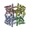
 Components
 Homo sapiens (human) / Gene: PDE4D2 / Plasmid: pET15b / Production host:
 X-RAY DIFFRACTION / Number of used crystals: 1
  Sample preparation
 SYNCHROTRON / Site:
  NSLS
NSLS  / Beamline: X25 / Wavelength: 1 Å
 Processing
 MOLECULAR REPLACEMENT
 Movie
Movie Controller



 PDBj
PDBj


