 Movie
Movie Controller

[English] 日本語
 Yorodumi
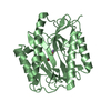
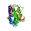
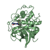
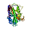
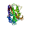
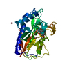
- PDB-2evo: crystal structure of methionine amino peptidase in complex with N... -
+ Open data

- Basic information
| Entry | Database: PDB / ID: 2evo | ||||||
|---|---|---|---|---|---|---|---|
| Title | crystal structure of methionine amino peptidase in complex with N-cyclopentyl-N-(thiazol-2-yl)oxalamide | ||||||
 Components Components | Methionine aminopeptidase | ||||||
 Keywords Keywords | HYDROLASE / methionine aminopeptidase / complex | ||||||
| Function / homology |  Function and homology information Function and homology informationmethionyl aminopeptidase / initiator methionyl aminopeptidase activity / metalloaminopeptidase activity / ferrous iron binding / proteolysis / cytosol Similarity search - Function | ||||||
| Biological species |  | ||||||
| Method |  X-RAY DIFFRACTION / MOLECULAR REPLACEMENT / Resolution: 1.7 Å X-RAY DIFFRACTION / MOLECULAR REPLACEMENT / Resolution: 1.7 Å | ||||||
 Authors Authors | Huang, W.-J. | ||||||
 Citation Citation | Journal: Acta Crystallogr.,Sect.D / Year: 2006 Title: Structural analysis of metalloform-selective inhibition of methionine aminopeptidase. Authors: Xie, S.X. / Huang, W.J. / Ma, Z.Q. / Huang, M. / Hanzlik, R.P. / Ye, Q.Z. | ||||||
| History |
|
- Structure visualization
| Structure viewer | Molecule: MolmilJmol/JSmol |
|---|
- Downloads & links
-Download
| PDBx/mmCIF format | 2evo.cif.gz | 119.9 KB | Display | PDBx/mmCIF format |
|---|---|---|---|---|
| PDB format | pdb2evo.ent.gz | 91.3 KB | Display | PDB format |
| PDBx/mmJSON format | 2evo.json.gz | Tree view | PDBx/mmJSON format | |
| Others |  Other downloads Other downloads |
-Validation report
| Arichive directory | https://data.pdbj.org/pub/pdb/validation_reports/ev/2evoftp://data.pdbj.org/pub/pdb/validation_reports/ev/2evo | HTTPS FTP |
|---|
-Related structure data
| Related structure data | 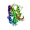 2evcC  2evmC 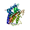 2matS C: citing same article ( S: Starting model for refinement |
|---|---|
| Similar structure data |


-Links
 PDBj
PDBj
- Assembly
| Deposited unit | 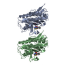
| ||||||||
|---|---|---|---|---|---|---|---|---|---|
| 1 | 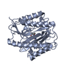
| ||||||||
| 2 | 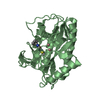
| ||||||||
| Unit cell |
|
-Components
| #1: Protein | Mass: 29370.838 Da / Num. of mol.: 2 Source method: isolated from a genetically manipulated source Source: (gene. exp.) #2: Chemical | ChemComp-CO /   Mass: 58.933 Da / Num. of mol.: 6 / Source method: obtained synthetically / Formula: Co Mass: 58.933 Da / Num. of mol.: 6 / Source method: obtained synthetically / Formula: Co#3: Chemical | 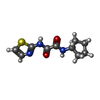  Mass: 239.294 Da / Num. of mol.: 2 / Source method: obtained synthetically / Formula: C10H13N3O2S Mass: 239.294 Da / Num. of mol.: 2 / Source method: obtained synthetically / Formula: C10H13N3O2S#4: Water | ChemComp-HOH / |  Mass: 18.015 Da / Num. of mol.: 263 / Source method: isolated from a natural source / Formula: H2O Mass: 18.015 Da / Num. of mol.: 263 / Source method: isolated from a natural source / Formula: H2O |
|---|
-Experimental details
-Experiment
| Experiment | Method: X-RAY DIFFRACTION / Number of used crystals: 1 |
|---|
- Sample preparation
| Crystal | Density Matthews: 1.9 Å3/Da / Density % sol: 35.1 % |
|---|---|
| Crystal grow | Temperature: 292 K / Method: vapor diffusion / pH: 7.5 Details: 15% PEG 8000, 0.1M HEPES, pH 7.5, VAPOR DIFFUSION, temperature 292K |
-Data collection
| Diffraction | Mean temperature: 100 K |
|---|---|
| Diffraction source | Source: ROTATING ANODE / Type: RIGAKU RUH3R / Wavelength: 1.5418 Å |
| Detector | Type: RIGAKU RAXIS IV / Detector: IMAGE PLATE / Date: Jun 7, 2004 / Details: mirrors |
| Radiation | Monochromator: GRAPHITE / Protocol: SINGLE WAVELENGTH / Monochromatic (M) / Laue (L): M / Scattering type: x-ray |
| Radiation wavelength | Wavelength: 1.5418 Å / Relative weight: 1 |
| Reflection | Resolution: 1.7→20 Å / Num. all: 43944 / Num. obs: 48408 / % possible obs: 92.1 % / Observed criterion σ(F): 0 / Observed criterion σ(I): 0 |
| Reflection shell | Resolution: 1.7→2.1 Å / % possible all: 89.3 |
- Processing
| Software |
| |||||||||||||||||||||
|---|---|---|---|---|---|---|---|---|---|---|---|---|---|---|---|---|---|---|---|---|---|---|
| Refinement | Method to determine structure: MOLECULAR REPLACEMENT Starting model: 2mat Resolution: 1.7→20 Å / σ(F): 0 / Stereochemistry target values: Engh & Huber
| |||||||||||||||||||||
| Refinement step | Cycle: LAST / Resolution: 1.7→20 Å
| |||||||||||||||||||||
| Refine LS restraints |
|
